Macular Morphological Changes of Persistent Subclinical Subretinal Fluid after Scleral Buckle Surgery:An Optical Coherence Tomography Study
-
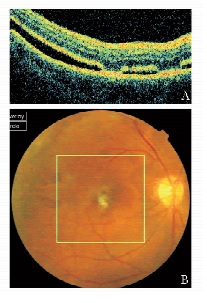
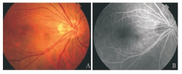
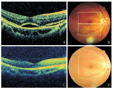
摘要:目的 探讨孔源性视网膜脱离术后亚临床性视网膜下液的黄斑区光学相干断层扫描(optical coherence tomo-graphy, OCT)表现。方法 对孔源性视网膜脱离巩膜扣带术后临床视网膜复位但视力恢复不佳或有视物变形症状患者19例21眼, 应用3D OCT在其黄斑部行范围为6 mm×6 mm×1.7 mm、分辨率为512×128的三维扫描检查。分析患者黄斑部的OCT表现, 包括中心凹神经上皮厚度、神经上皮脱离情况、感光细胞内外节(inner segment/outer segment, IS/OS)结合部情况等。结果 3D OCT检查显示15眼存在黄斑区亚临床性视网膜下液, 均为术前黄斑脱离的患者。其中14眼神经上皮的IS/OS结合部反光信号完整且增厚增强, 2眼可见中心凹外色素上皮层的高反射隆起, 2眼可见视网膜前膜反射信号。15眼中, 7眼神经上皮断续脱离。7例患者进行了多次OCT随访, 时间为术后3~12个月, 神经上皮脱离最终全部消失, 最佳矫正视力恢复到0.7~1.2。结论 3D OCT能发现临床视网膜复位患者的亚临床性视网膜下液并确定脱离范围, 大多数患者表现为神经上皮浅脱离, IS/OS结合部反光信号完整且增厚增强。
-
关键词:
- 孔源性视网膜脱离 /
- 谱域光学相干断层扫描 /
- 巩膜扣带术 /
- 亚临床性视网膜下液
Abstract:Objective To evaluate macular morphological changes by spectral domain optical coherence tomography (3D OCT) in patients with persistent subclinical subretinal fluid after successful scleral buckle surgeries.Methods Totally 19 cases (21 eyes) with incomplete visual acuity recovery or metamorphosis were reviewed. 3D OCT was performed in the macula (size:6 mm×6 mm×1.7 mm; resolution:512×128). The morphological changes of macula including thickness of foveal neuroepithelium, neuroepithelial detachment, inner segment/outer segment (IS/OS) junction were recorded.Results 3D OCT showed persistent subclinical subretinal fluid in 15 eyes, and all of them occurred in patients with preoperative macular detachment. Also, 14 of 15 eyes showed disruption of the photoreceptor IS/OS junction, 2 showed irregularity of retinal pigment epithelium reflection signal, and 2 showed thin epiretinal membrane. Seven of these 15 eyes showed discontinuous retinal detachment. During the 3to 12month continuous follow-up for 7 eyes, the neuroepithelial detachment finally disappeared, and the best corrected visual acuity returned to 0.7-1.2.Conclusions 3D OCT can demonstrate areas of persistent subclinical subretinal fluid after successful scleral buckle surgeries. Most patients have neural epithelium shallow detachment as well as complete and enhanced IS/OS junction signal. -
2019新型冠状病毒(2019 novel coronavirus, 2019-nCoV)属于β属冠状病毒,2019年12月以来我国陆续出现该病毒感染引起的以肺部病变为主的新型传染病。临床特征为发热、干咳、气促、外周血白细胞一般不高/降低、胸部X线检查显示炎症性改变等。我国已将2019-nCoV感染的肺炎定为法定传染病。为规范诊疗,北京协和医院特成立专家组并制订2019-nCoV感染的肺炎诊疗方案(V2.0)。
1. 医务人员的防护
1.1 接诊医护准入
一线医护人员应进行上岗前筛查和2019-nCoV知识培训,并需排除以下情况:孕妇、年龄超过55岁、慢性疾病史(慢性肝炎、慢性肾炎、糖尿病、自身免疫性疾病及肿瘤)、合并急性发热者。
上岗前筛查血常规、尿常规、生化、肌酸激酶,并进行胸部X线检查。
1.2 隔离和防护要求
参见国家卫生健康委员会发布的“新型冠状病毒感染的肺炎诊疗方案(试行第三版)”[1]。
1.3 2019-nCoV密切接触后医护人员的隔离观察
(1) 与2019-nCoV感染的肺炎患者密切接触的医护人员应相对隔离,避免到处走动,避免广泛接触。
(2) 出现发热、咳嗽、气短等症状时应立即隔离,并进行相关检查。
(3) 结束2019-nCoV感染的肺炎病区工作时,应进行咽拭子及血常规检查,有异常者应接受严格隔离观察;无异常者普通隔离观察1周后恢复工作。
2. 2019-nCoV感染患者的诊疗
2.1 筛查标准[1-2]
(1) 流行病学史:发病前2周内有湖北旅行或居住史,或发病前14 d内曾接触过来自湖北、发热且伴有呼吸道症状的患者,或有聚集性发病。
(2) 72 h以内的急性发热,不伴流感样症状,且未证实其他病因者。
2.2 诊断标准
(1) 流行病学史。
(2) 临床表现:发热;发病早期白细胞总数正常/降低,或淋巴细胞计数减少;胸部影像学早期呈现多发小斑片影及间质改变,以肺外带明显, 进而发展为双肺多发磨玻璃影、浸润影,严重者可出现肺实变。
(3) 确诊:痰液、咽拭子、下呼吸道分泌物等标本行实时荧光RT-PCR检测2019-nCoV核酸阳性。
(4) 对于急性发热(72 h内,体温>37.5 ℃)且肺部影像学正常者,若外周血淋巴细胞绝对值<0.8×10⁹/L,或出现CD4+及CD8+T细胞计数明显下降者,即使核酸检测未呈现阳性,均应居家隔离密切观察;必要时可考虑24 h后复查核酸检测,并依据临床表现复查胸部CT。
2.3 2019-nCoV感染患者检查常规
2.3.1 筛查病例就诊当天
应进行痰/咽拭子的核酸检查、血常规、尿常规、血气、肝肾功能、C-反应蛋白、降钙素原、肌酸激酶+肌红蛋白、凝血及胸部CT。可酌情查炎症因子[如白细胞介素(interleukin, IL)-6、IL-10、肿瘤坏死因子-α]、TB淋巴细胞亚群11项、补体[3-5]。
2.3.2 确诊患者序贯检查
(1) 留观后第3、5、7天及出院时依据病情,可检查血细胞、肝肾功能、肌酸激酶+肌红蛋白、凝血、C-反应蛋白;第5~7天若有条件可复查降钙素原及TB淋巴细胞亚群11项[3-5]。
(2) 留观后1~2 d均需复查胸部X线,之后视病情决定,但复查时间不超过5 d。
(3) 非转院患者出院前应复查血常规、胸部X线、肝肾功能及入院时所有异常检查。
2.4 根据病情严重程度确定治疗场所
所有具有筛查指征的病例均需就地医学隔离(单间隔离),一旦确诊均需前往指定医院救治。
2.4.1 重症病例
按照国家卫生健康委员会定义[1], 符合如下标准之一,需留院治疗并尽快转运至北京市定点诊治医疗机构: (1)呼吸频率增快(≥30次/min),呼吸困难;(2)吸空气时指氧饱和度≤95%,或动脉血氧分压/吸氧浓度≤300 mmHg(1 mmHg=0.133 kPa);(3)肺部影像学显示多叶病变或48 h内病灶进展>50%;(4)快速序贯性器官功能衰竭评分(quick sequential organ failure assessment,qSOFA)≥2分;(5)社区获得性肺炎CURB-65评分≥1分;(6)合并气胸;(7)需住院治疗的其他临床情况。
2.4.2 危重症病例
按照国家卫生健康委员会定义[1], 符合呼吸衰竭、感染性休克、合并其他器官功能衰竭标准之一,立即进入ICU并在条件允许时尽快转运至定点医疗机构诊治。
2.5 治疗
2.5.1 一般治疗
卧床休息,监测生命体征、指氧饱和度,支持对症治疗,保证热量,维持水、电解质及酸碱平衡等内环境稳定。
2.5.2 氧疗
存在低氧血症者立即进行氧疗,血氧饱和度维持目标:非妊娠患者≥90%,妊娠患者92%~ 95%。
2.5.2.1 氧疗方式
轻症患者初始给予普通鼻导管吸氧,以5 L/min开始;重症患者如呼吸窘迫加重或标准氧疗无效时,可给予高流量鼻导管吸氧,以20 L/min起始,逐步上调至50~60 L/min,同时依据氧合目标调整吸氧浓度。
2.5.2.2 呼吸支持方式
无创呼吸机仅在患者可很好耐受无创通气时使用,不建议先于高流量鼻导管吸氧使用;有创呼吸机气管插管应由经验丰富人员完成,操作时按全面防护要求进行,呼吸机设置遵循急性呼吸窘迫综合征保护性通气策略进行;当有创呼吸机无法维持氧合时,可给予俯卧位体外膜肺氧合相应治疗,由于该操作复杂,需同时注意全面防护以及预防院内感染。
2.5.3 抗病毒治疗
目前尚无循证医学证据支持现有抗病毒药物对2019-nCoV有效,可酌情用洛匹那韦/利托那韦每次2粒×2次/d,疗程14 d。
2.5.4 糖皮质激素
重症患者酌情早期使用糖皮质激素,静脉滴注甲泼尼龙40~80 mg×1次/d,疗程5 d;可根据患者临床病情及影像学表现酌情延长疗程。
2.5.5 人免疫球蛋白
重症患者依据病情可酌情早期静脉输注人免疫球蛋白0.25~0.50 g/ (kg·d),疗程3~5 d[6-7]。
2.5.6 经验性抗菌治疗
根据患者临床和影像学表现,如不能除外合并细菌感染,轻症患者可口服针对社区获得性肺炎的抗菌药物,如二代头孢或氟喹诺酮类;重症患者需覆盖所有可能的病原体。
3. 防护和转运
(1) 重症患者一旦确诊,且有气管插管风险,应立即转运至有负压条件的ICU病房进行治疗;操作时按全面防护要求进行。
(2) 转运途中使用储氧面罩15 L/min以上给氧,保证储氧气囊充气满意。
(3) 气管插管应使用标准快速顺序诱导插管,尽可能使用肌松药物,最大程度避免患者呛咳引起飞沫传播。
(4) 插管后的眼罩等重复使用物品应使用健之素消毒后方可拿出负压病房。
(5) 插管患者应使用密闭吸痰器吸痰,避免呼吸机气流引起空气传播。
(6) 特殊情况下必须断开呼吸机进行气道操作时,应使用呼吸机的待机功能,避免呼吸机气流引起空气传播。如呼吸机无待机功能,应阻断呼吸机Y型管口,避免空气播散。
4. 解除隔离和出院标准[1]
体温恢复正常3 d以上,呼吸道症状明显好转,肺部影像学炎症明显吸收,且连续两次呼吸道病原核酸检测阴性(采样时间间隔至少1 d),可解除隔离出院或根据病情转至相应科室治疗其他疾病。
执笔人
李太生 (北京协和医院感染内科)
曹玮 (北京协和医院感染内科)
翁利 (北京协和医院内科ICU)
范洪伟 (北京协和医院感染内科)
施举红 (北京协和医院呼吸与危重症医学科)
参与讨论专家 (按姓氏汉语拼音排序)
柴文昭 杜斌 郭娜 韩丁 韩扬 胡小芸
焦洋 金征宇 刘正印 隆云 马小军 潘慧
王惠珍 王孟昭 吴文铭 吴欣娟 徐英春 许文兵
张波 张奉春 张圣洁 朱华栋
-
表 1 多次光学相干断层扫描随访患者的亚临床性视网膜下液及随诊情况

-
[1] Kaga T, Fonseca RA, Dantas MA, et al. Optical coherence tomography of bleb-like subretinal lesions after retinal reattachment surgery[J]. Am J Ophthalmol, 2001, 132:120-121. DOI: 10.1016/S0002-9394(00)00950-8
[2] Wolfensberger TJ, Gonvers M. Optical coherence tomography in the evaluation of incomplete visual acuity recovery after macula-off retinal detachments[J]. Graefes Arch Clin Exp Ophthalmol, 2002, 240:85-89. DOI: 10.1007/s00417-001-0410-6
[3] Gharbiya M, Grandinetti F, Scavella V, et al. Correlation between spectral-domain optical coherence tomography findings and visual outcome after primary rhegmatogenous retinal detachment repair[J]. Retina, 2012, 32:43-53. DOI: 10.1097/IAE.0b013e3182180114
[4] Rossetti A, Doro D, Manfrè A, et al. Long-term follow-up with optical coherence tomography and microperimetry in eyes with metamorphopsia after macula-off retinal detachment repair[J]. Eye (Lond), 2010, 24:1808-1813. DOI: 10.1038/eye.2010.138
[5] Cavallini GM, Masini C, Volante V, et al. Visual recovery after scleral buckling for macula-off retinal detachments:an optical coherence tomography study[J]. Eur J Ophthalmol, 2007, 17:790-796. DOI: 10.1177/112067210701700517
[6] Veckeneer M, Derycke L, Lindstedt EW, et al. Persistent subretinal fluid after surgery for rhegmatogenous retinal detachment:hypothesis and review[J]. Graefes Arch Clin Exp Ophthalmol, 2012, 250:795-802. DOI: 10.1007/s00417-011-1870-y
[7] Seo JH, Woo SJ, Park KH, et al. Influence of persistent submacular fluid on visual outcome after successful scleral buckle surgery for macula-off retinal detachment[J]. Am J Ophthalmol, 2008, 145:915-922. DOI: 10.1016/j.ajo.2008.01.005
[8] Smith AJ, Telander DG, Zawadzki RJ, et al. High-resolution Fourier-domain optical coherence tomography and microperimetric findings after macula-off retinal detachment repair[J]. Ophthalmology, 2008, 115:1923-1929. DOI: 10.1016/j.ophtha.2008.05.025
[9] Feraoun MN, Dot C, Lecorre A, et al. Delayed subretinal fluid absorption after rhegmatogenous retinal detachment[J]. J Fr Ophtalmol, 2011, 34:248-251. DOI: 10.1016/j.jfo.2011.01.006
[10] Benson SE, Schlottmann PG, Bunce C, et al. Optical coherence tomography analysis of the macula after vitrectomy surgery for retinal detachment[J]. Ophthalmology, 2006, 113:1179-1183. DOI: 10.1016/j.ophtha.2006.01.039
[11] Wakabayashi T, Oshima Y, Fujimoto H, et al. Foveal microstructure and visual acuity after retinal detachment repair:imaging analysis by Fourier-domain optical coherence tomography[J]. Ophthalmology, 2009, 116:519-528. DOI: 10.1016/j.ophtha.2008.10.001
[12] Wolfensberger TJ. Foveal reattachment after macula-off retinal detachment occurs faster after vitrectomy than after buckle surgery[J]. Ophthalmology, 2004, 111:1340-1343. DOI: 10.1016/j.ophtha.2003.12.049
[13] 姚进, 沈轶, 蒋沁.视网膜脱离术前和术后黄斑区改变的光学相干断层扫描评估[J].南京医科大学学报:自然科学版, 2007, 27:974-976. http://www.wanfangdata.com.cn/details/detail.do?_type=perio&id=njykdxxb200709018 -
期刊类型引用(2)
1. 彭珊珊,龚健杨,李琳娜. 视网膜脱离巩膜扣带术后持续性视网膜下积液与视力的关系. 中国医学工程. 2022(07): 34-38 .  百度学术
百度学术
2. 陈力菲,于旭辉. 孔源性视网膜脱离巩膜扣带术后持续性视网膜下液的研究进展. 国际眼科杂志. 2018(07): 1237-1240 .  百度学术
百度学术
其他类型引用(10)

 作者投稿
作者投稿 专家审稿
专家审稿 编辑办公
编辑办公 邮件订阅
邮件订阅 RSS
RSS


 下载:
下载:


