 作者投稿
作者投稿 专家审稿
专家审稿 编辑办公
编辑办公 邮件订阅
邮件订阅 RSS
RSS

Prevention of Postoperative Adhesions with N, O-carboxymethyl Chitosan in Rabbit: A Large-sample Gross and Histopathological Observation
-
摘要:
目的 评价羧甲基壳聚糖(N, O-carboxymethyl chitosan, NOCC)预防兔手术后粘连的效果。 方法 220只雌性大耳白兔采用双子宫模型造成手术粘连, 随机分组后接受单纯造模手术(对照组)或造模手术+关腹前腹腔内注射NOCC(NOCC组)。所有手术均由同一位术者完成。每组各22只大耳白兔在实验设计的5个观察时间点(术后3、7、14、28和42 d)分别被处死并评估其粘连情况, 包括范围、类型和强度等大体情况和炎症、纤维化以及新生血管等镜下情况。 结果 NOCC组在术后3 d时其粘连的范围(P=0.0337)和强度(P=0.0271)以及炎症反应情况(P < 0.0001)均显著轻于对照组。在术后14 d内, NOCC组的纤维化情况均明显轻于对照组(P均 < 0.0005)。与对照组相比, NOCC组的粘连强度在术后14、28、42 d均明显减轻(P均 < 0.05), 而其粘连类型评分在术后28和42 d也明显降低(P均 < 0.05)。 结论 经NOCC处理后, 兔盆腹腔术后粘连大体和病理学评分均不同程度降低。应用NOCC可有效预防盆腹腔术后粘连。 Abstract:Objective To assess the effect of N, O-carboxymethyl chitosan (NOCC) in preventing postoperative adhesion in a rabbit model. Methods Double uterine horn model was established in 220 female rabbits to induce postoperative adhesion. The rabbits were randomized to receive either adhesion-inducing operation only (control group) or adhesion-inducing operation + intraperitoneal injection of NOCC before closure (NOCC group). All the operations were performed by one operator. Twenty-two rabbits from each group were euthanized at one of the five different time points (postoperative day 3, 7, 14, 28, and 42), and adhesion formation was scored both grossly (extent, type, and tenacity) and histopathologically (inflammation, fibrosis, and vascularization). Results The extent (P=0.0337) and tenacity of adhesion (P=0.0271) as well as inflammation(P < 0.0001) were lower in the NOCC group than in the control group on day 3. Fibrosis was less obvious in the NOCC group compared to the control group (P < 0.0005) before day 14. The tenacity scores of adhesion on day 14, 28, and 42 were significantly lower in the NOCC group than in the control group(all P < 0.05), while the type scores were obviously lower in the NOCC group on day 28 and 42(all P < 0.05). Conclusions Treatment with NOCC could reduce both gross and histopathological scores of surgery-induced abdominopelvic adhesions in rabbits. NOCC could be an effective therapy for preventing postoperative abdominopelvic adhesion. -

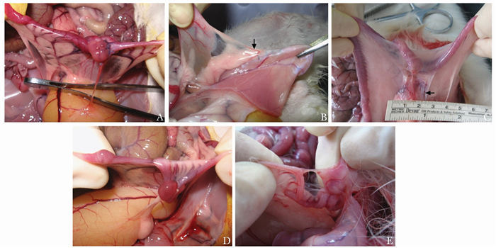
图 1 大耳白兔双子宫造模手术后盆腹腔粘连的大体观察
因两组变化趋势相似,故A~D均展示对照组大体所见;A.术后3 d对照组:子宫和膀胱间的疏松、膜状粘连;B.术后7 d对照组:可见1例新生血管(箭头);C.术后28 d对照组:新生血管网形成(箭头);D.术后42 d对照组:粘连范围较28 d降低;E.术后42 d NOCC组:粘连更疏松,新生血管更细NOCC:羧甲基壳聚糖

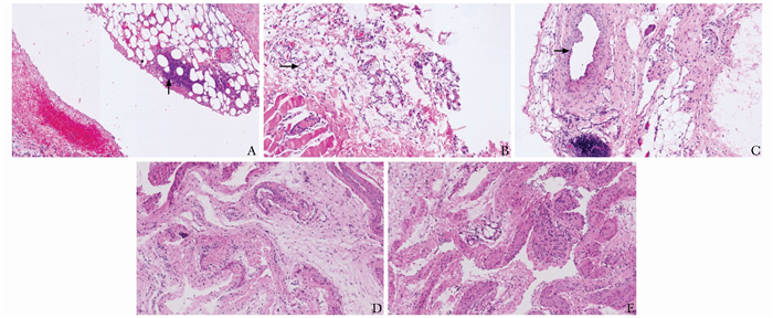
图 2 大耳白兔双子宫造模手术后盆腹腔粘连的病理表现(HE染色,×150)
A.术后3 d对照组:可见微脓肿(箭头);B.术后7 d对照组:可见新生微毛细管(箭头);C.术后28 d对照组:可见一条有完整结构的大毛细血管,在其下方可见慢性炎症细胞浸润(箭头);D.术后42 d对照组:纤维密度增加; E.术后42 d NOCC组:新生血管与术后28 d相似NOCC:同图 1
表 1 NOCC组和对照组在术后5个时间点的大体粘连评价情况
术后时间(d) 粘连特征 对照组[M(Q1, Q3)] NOCC组[M(Q1, Q3)] P 3 范围 2 (1,2) 1 (1,2) 0.0337 类型 2 (2,2) 2 (2,2) 0.7638 强度 2 (1.25,2) 1 (1,2) 0.0271 7 范围 2 (1,2) 2 (1,2) 0.4257 类型 2 (2,2) 2 (1.25,2) 0.4609 强度 2 (1.25,2) 1 (1,2) 0.1026 14 范围 2 (1,2) 1.5(1,2) 0.4693 类型 2 (2,1) 2 (1,2) 0.1142 强度 2 (1,2.75) 1 (1,2) 0.0410 28 范围 2.5(1,2) 2 (1.25,2) 0.5006 类型 2 (2,2) 2 (1,2) 0.0210 强度 2 (2,2) 1 (1,1) <0.0001 42 范围 2 (1,2) 1 (0,1) 0.1475 类型 2 (1,2) 1 (0,1) 0.0027 强度 2 (1,2) 1 (0,1) 0.0002 NOCC:同图 1  下载: 导出CSV
表 2 NOCC组和对照组在术后5个时间点的镜下粘连评价情况
术后时间(d) 镜下所见 对照组[M(Q1, Q3)] NOCC组[M(Q1, Q3)] P 3 炎症反应 5 (4, 6) 3(1, 3.75) <0.0001 纤维化 4 (3, 4) 3(2, 3) 0.0004 血管增生 0 (0, 0) 0(0, 0) 1.0000 7 炎症反应 2 (1, 3) 2(0, 4) 0.9044 纤维化 3 (3, 3.75) 1(1, 2) <0.0001 血管增生 2 (2, 4.75) 2(2, 3) 0.2717 14 炎症反应 1 (0.25, 2) 1(0, 1) 0.8226 纤维化 3 (3, 4) 2(0.25, 2.75) 0.0002 血管增生 4 (1.25, 5) 2(1, 3.75) 0.1012 28 炎症反应 0 (0, 1) 1(0, 2) 0.1468 纤维化 3 (3, 3.75) 3(2, 3) 0.4036 血管增生 2.5(2, 4) 3(2, 3) 0.8155 42 炎症反应 0 (0, 1) 1(0, 1) 0.4109 纤维化 3 (2.25, 4) 2(2, 3) 0.0501 血管增生 4 (2, 4.75) 3(2, 3) 0.2626 NOCC:同图 1
下载: 导出CSV
-
[1] Stanciu D, Menzies D. The magnitude of adhesion-related problems[J]. Colorectal Dis, 2007, 9:35-38. doi: 10.1111/j.1463-1318.2007.01346.x [2] Liakakos T, Thomakos N, Fine PM, et al. Peritoneal adhesions:etiology, pathophysiology, and clinical significance. Recent advances in prevention and management[J]. Dig Surg, 2001, 18:260-273. doi: 10.1159/000050149 [3] Wiseman DM. Disorders of adhesions or adhesion-related disorder:monolithic entities or part of something bigger-CAPPS?[J]. Semin Reprod Med, 2008, 26:356-368. doi: 10.1055/s-0028-1082394 [4] Ten Broek RP, Kok-Krant N, Bakkum EA, et al. Different surgical techniques to reduce post-operative adhesion formation:a systematic review and meta-analysis[J]. Hum Reprod Update, 2013, 19:12-25. doi: 10.1093/humupd/dms032 [5] Sahbaz A, Aynioglu O, Isik H, et al. Bromelain:a natural proteolytic for intra-abdominal adhesion prevention[J]. Int J Surg, 2015, 14:7-11. doi: 10.1016/j.ijsu.2014.12.024 [6] Moraloglu O, Işik H, Kilic S, et al. Effect of bevacizumab on postoperative adhesion formation in a rat uterine horn adhesion model and the correlation with vascular endothelial growth factor and Ki-67 immunopositivity[J]. Fertil Steril, 2011, 95:2638-2641. doi: 10.1016/j.fertnstert.2011.02.005 [7] Sakai S, Ueda K, Taya M. Peritoneal adhesion prevention by a biodegradable hyaluronic acid-based hydrogel formed in situ through a cascade enzyme reaction initiated by contact with body fluid on tissue surfaces[J]. Acta Biomater, 2015, 24:152-158. doi: 10.1016/j.actbio.2015.06.023 [8] Hellebrekers BW, Trimbos-Kemper GC, van Blitterswijk CA, et al. Effects of five different barrier materials on postsurgical adhesion formation in the rat[J]. Hum Reprod, 2000, 15:1358-1363. doi: 10.1093/humrep/15.6.1358 [9] Ong SY, Wu J, Moochhala SM, et al. Development of a chitosan based wound dressing with improved hemostatic and antimicrobial properties[J]. Biomaterials, 2008, 29:4323-4332. doi: 10.1016/j.biomaterials.2008.07.034 [10] Rabea EI, Badawy ME, Stevens CV, et al. Chitosan as antimicrobial agent:Applications and mode of action[J]. Biomacromolecules, 2003, 4:1457-1465. doi: 10.1021/bm034130m [11] Krause TJ, Goldsmith NK, Ebner S, et al. An inhibitor of cell proliferation associated with adhesion formation is suppressed by N, O-carboxymethyl chitosan[J]. J Invest Surg, 1998, 11:105-113. doi: 10.3109/08941939809032189 [12] Zhou J, Liwski RS, Elson C, et al. Reduction in postsurgical adhesion formation after cardiac surgery in a rabbit model using N, O-carboxymethyl chitosan to block cell adherence[J]. J Thorac Cardiovasc Surg, 2008, 135:777-783. doi: 10.1016/j.jtcvs.2007.09.033 [13] Ren C, Zhu L, Sun JC. Creation of an animal model for post-operative adhesion prevention[J]. Zhongguo Yi Xue Ke Xue Yuan Xue Bao, 2012, 34:109-115. http://www.wanfangdata.com.cn/details/detail.do?_type=perio&id=zgyxkxyxb201202003 [14] Adhesion Scoring Group. Improvement of interobserver reproducibility of adhesion scoring systems[J]. Fertil Steril, 1994, 62:984-988. doi: 10.1016/S0015-0282(16)57062-9 [15] American Fertility Society. The American Fertility Society classification of adnexal adhesions, distal tubal occlusion, tubal occlusion secondary to tubal ligation, tubal pregnancies, Mullerian anomalies and intrauterine adhesions[J]. Fertil Steril, 1988, 49:944-955. doi: 10.1016/S0015-0282(16)59942-7 [16] Ersoy E, Ozturk V, Yazgan A, et al. Comparison of the two types of bioresorbable barriers to prevent intra-abdominal adhesions in rats[J]. J Gastrointest Surg, 2009, 13:282-286. doi: 10.1007/s11605-008-0678-5 [17] Hooker GD, Taylor BM, Driman DK. Prevention of adhesion formation with use of sodium hyaluronate-based bioresorbable membrane in a rat model of ventral hernia repair with polypropylene mesh-A randomised, controlled study[J]. Surgery, 1999, 125:211-216. doi: 10.1016/S0039-6060(99)70267-9 [18] Ahmad G, Mackie FL, Iles DA, et al. Fluid and pharmacological agents for adhesion prevention after gynaecological surgery[J]. Cochrane Database Syst Rev, 2014, 7:CD001298. [19] Costain DJ, Kennedy R, Ciona C, et al. Prevention of post-surgical adhesions with N, O-carboxymethyl chitosan:examination of the most efficacious preparation and the effect of N, O-carboxymethyl chitosan on postsurgical healing[J]. Surgery, 1997, 121:314-319. doi: 10.1016/S0039-6060(97)90360-3 [20] Zhou J, Elson C, Lee TDG. Reduction in postoperative adhesion formation and re-formation after an abdominal operation with the use of N, O-carboxymethyl chitosan[J]. Surgery, 2004, 135:307-312. doi: 10.1016/j.surg.2003.07.005 [21] Wang D, Mo J, Pan S, et al. Prevention of postoperative peri-toneal adhesions by O-carboxymethyl chitosan in a rat cecal abrasion mode[J]. Clin Invest Med, 2010, 33:E254-E260. doi: 10.25011/cim.v33i4.14228 [22] Ricketts SA, Sibbons PD, Green CJ. Quantitative analysis of the development of experimentally induced post surgical adhesions:a microstereological study[J]. Int J Exp Pathol, 1999, 80:325-334. [23] Harris ES, Morgan RF, Rodeheaver GT. Analysis of the kinetics of peritoneal adhesion formation in the rat and evaluation of potential antiadhesive agents[J]. Surgery, 1995, 117:663-669. doi: 10.1016/S0039-6060(95)80010-7 [24] Yeo Y, Highley CB, Bellas E, et al. In situ cross-linkable hyaluronic acid hydrogels prevent post-operative abdominal adhesions in a rabbit model[J]. Biomaterials, 2006, 27:4698-4705. doi: 10.1016/j.biomaterials.2006.04.043 [25] Li XD, Xia DL, Shen LL, et al. Effect of "phase change" complex on postoperative adhesion prevention[J]. J Surg Res, 2016, 202:216-224. doi: 10.1016/j.jss.2015.12.033 [26] Haney AF, Doty E. The formation of coalescing peritoneal adhesions requires injury to both contacting peritoneal surfaces[J]. Fertil Steril, 1994, 61:767-775. doi: 10.1016/S0015-0282(16)56660-6 [27] van Hinsbergh VW, Kooistra T, Scheffer MA, et al. Characterization and fibrinolytic properties of human omental tissue mesothelial cells. Comparison with endothelial cells[J]. Blood, 1990, 75:1490-1497. doi: 10.1182/blood.V75.7.1490.1490 [28] Hickey MJ. Role of inducible nitric oxide synthase in the re-gulation of leukocyte recruitment[J]. Clin Sci, 2001, 100:1-12. [29] Milligan DW, Raftery AT. Observations on the pathogenesis of peritoneal adhesions:a light and electron microscopical study[J]. Br J Surg, 1974, 61:274-280. doi: 10.1002/bjs.1800610406 [30] Duran B, Ak D, Cetin A, et al. Reduction of postoperative adhesions by N, O-carboxymethyl chitosan and spermine NONOate in rats[J]. Exp Anim, 2003, 52:267-272. doi: 10.1538/expanim.52.267 [31] Cetin M, Ak D, Duran B, et al. Use of methylene blue and N, O-carboxymethyl chitosan to prevent postoperative adhesions in a rat uterine horn model[J]. Fertil Steril, 2003, 80 Suppl 2:698-701. [32] Diamond MP, Luciano A, Johns DA, et al. Reduction of postoperative adhesions by N, O-carboxymethyl chitosan:a pilot study[J]. Fertil Steril, 2003, 80:631-636. doi: 10.1016/S0015-0282(03)00759-3 -

 点击查看大图
图(2) / 表(2)
计量
- 文章访问数: 117
- HTML全文浏览量: 26
- PDF下载量: 6
- 被引次数: 0


