 作者投稿
作者投稿 专家审稿
专家审稿 编辑办公
编辑办公 邮件订阅
邮件订阅 RSS
RSS

-
摘要:
目的 研究超声三血管气管切面对于胎儿主动脉弓异常的诊断价值。 方法 回顾性分析2010年5月至2014年5月北京协和医院诊断的17例胎儿主动脉弓异常的产前超声图像资料, 并与产后超声及病理结果进行对照, 分析不同类型主动脉弓异常的产前超声特征, 重点关注三血管气管切面的表现。 结果 17例主动脉弓异常在三血管气管切面均有阳性表现。其中主动脉弓缩窄3例, 表现为主动脉弓细小; 主动脉弓离断7例, 表现为主动脉变细, 不能连接至降主动脉; 右位主动脉弓合并迷走左锁骨下动脉5例, 可见环绕气管的"U"形血管环; 右位主动脉弓合并镜像分支2例, 可见主动脉弓位于气管右侧, 动脉导管不汇入降主动脉。 结论 三血管气管切面是提示胎儿主动脉弓异常敏感且有效的切面, 中孕超声筛查时应重视该切面, 早期发现异常并给予适当的遗传咨询。 Abstract:Objective To investigate the diagnostic value of three-vessel and tracheal (3VT) view in fetal aortic arch anomalies. Methods Ultrasound findings of 17 fetuses with aortic arch anomalies diagnosed in Peking Union Medical College Hospital between May 2010 and May 2014 were reviewed and compared with the findings of postnatal ultrasonography and pathological examinations to analyze the ultrasound features of aortic arch anomalies of different types, focusing on the importance of 3VT view. Results In all 17 cases the 3VT view was abnormal. 3 cases were aortic coarctation, mainly manifested as reduced diameter of the aortic arch; 7 cases were interrupted aortic arch, in which there was discontinuity between the thinning aortic arch and the descending aorta; 5 cases were right aortic arch (RAA) with aberrant left subclavian artery, with a 'U'-shaped vascular loop around the trachea; and 2 cases were RAA with mirror-image branching, with the aortic arch situated to the right of the trachea and not forming a V- or U-shaped confluence with the ductus. Conclusions The 3VT view is avery sensitive view to effectively diagnose fetal aortic arch anomalies. Meticulous ultrasound examination focusing on this view during second trimester may disclose the presence of such anomalies and help provide the appropriate genetic counselling. -
Key words:
- ultrasonography /
- fetus /
- aortic arch anomalies /
- three-vessel and tracheal view
-

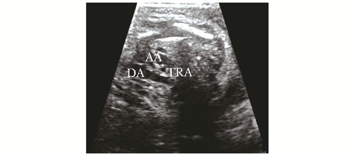
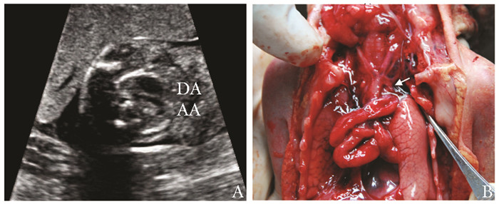
图 4 例17,孕23周,三血管气管切面(A)见主动脉弓位于气管右侧,动脉导管较细,位于气管左侧;左室流出道切面(B)可见主动脉增宽,骑跨于室间隔,为法洛四联症
AO:主动脉;PA:肺动脉;TRA:同图 3
表 1 17例胎儿主动脉弓异常的产前超声表现及妊娠结局
病例 孕妇年龄(岁) 月经孕周(周) 畸形类型 心脏超声各标准切面表现 合并心内异常 合并心外异常 妊娠结局 4CV LVOT RVOT 3VT AA 1 32 23 CoA + - - + ± RDH - 引产,未行病理 2 31 23 CoA + - - + - VSD 0.20 cm SUA、IUGR 引产,病理证实 3 22 24 CoA - - - + - PLSVC - 出生后无明显异常 4 31 24 IAA + + + + + VSD 0.39 cm、cTGA - 引产,病理为CoA 5 19 27 IAA - + - + + VSD 0.50 cm - 引产,未行病理 6 31 26 IAA + - - + + VSD 0.50 cm - 引产,病理证实 7 30 23 IAA + - - + + VSD 0.50 cm、RDH - 引产,病理证实 8 24 24 IAA + - - + + VSD 0.50 cm - 引产,未行病理 9 27 24 IAA - - - + + PLSVC - 引产,未行病理 10 30 25 IAA - - - + ± - - 引产,未行病理 11 27 24 RAA/ALSA - - - + - - - 2岁10个月,正常 12 32 24 RAA/ALSA - - - + - - - 2岁11个月,正常 13 33 23 RAA/ALSA - - - + - - - 2个月,正常 14 29 37 RAA/ALSA - - - + - - IUGR 2岁9个月,正常 15 29 27 RAA/ALSA - - - + - - - 2岁4个月,正常 16 24 21 RAA/镜像分支 + + + + - VSD 0.4 cm、DVOT - 引产,病理证实 17 26 23 RAA/镜像分支 + + + + - VSD 0.4 cm、TF - 引产,未行病理 CoA:主动脉弓缩窄;IAA:主动脉弓离断;RAA:右位主动脉弓;ALSA:迷走左锁骨下动脉;4CV:四腔心切面;LVOT:左室流出道切面;RVOT:右室流出道切面;3VT:三血管气管切面;AA:主动脉弓长轴切面;+:切面异常;-:切面正常;±:切面可疑;RDH:右心增大;VSD:室间隔缺损;PLSVC:永存左上腔静脉;cTGA:完全性大动脉转位;DVOT:右室双出口;TF:法洛四联症;SUA:单脐动脉;IUGR:宫内发育迟缓  下载: 导出CSV
-
[1] Yoo SJ, Min JY, Lee YH, et al. Fetal sonographic diagnosis of aortic arch anomalies[J]. Ultrasound Obstet Gynecol, 2003, 22:535-546. doi: 10.1002/uog.897 [2] 李胜利, 欧阳淑媛, 姚远, 等.先天性主动脉弓及其分支畸形产前超声诊断及围产期结局[J].中华医学超声杂志:电子版, 2011, 8:1675-1689. http://www.wanfangdata.com.cn/details/detail.do?_type=perio&id=zhyxcszz201108007 [3] 樊宜珍, 刘静华, 郑洪平, 等.胎儿主动脉弓异常的超声诊断和畸形特征分析[J].中国医药导刊, 2013, 15:1621-1622. http://www.wanfangdata.com.cn/details/detail.do?_type=perio&id=zgyydk201310038 [4] Gardiner H, Chaoui R. The fetal three-vessel and tracheal view revisited[J]. Semin Fetal Neonatal Med, 2013, 18:261-268. doi: 10.1016/j.siny.2013.01.007 [5] Kenny D, Hijazi ZM. Coarctation of the aorta: from fetal life to adulthood[J]. Cardiol J, 2011, 18:487-495. http://cn.bing.com/academic/profile?id=3aeaf28daf54bda1baf9a6bcae976f26&encoded=0&v=paper_preview&mkt=zh-cn [6] Celoria GC, Patton RB. Congenital absence of the aortic arch[J]. Am Heart J, 1959, 58:407-413. doi: 10.1016/0002-8703(59)90157-7 [7] Kanne JP, Godwin JD. Right aortic arch and its variants[J]. J Cardiovasc Comput Tomogr, 2010, 4:293-300. doi: 10.1016/j.jcct.2010.07.002 [8] Yagel S, Arbel R, Anteby EY, et al. The three vessels and trachea view (3VT) in fetal cardiac scanning[J]. Ultrasound Obstet Gynecol, 2002, 20: 340-345. doi: 10.1046/j.1469-0705.2002.00801.x [9] Jowett V, Aparicio P, Santhakumaran S, et al. Sonographic predictors of surgery in fetal coarctation of the aorta[J]. Ultrasound Obstet Gynecol, 2012, 40:47-54. http://cn.bing.com/academic/profile?id=0050f3072d45db6d29b4d2ea6ca7359e&encoded=0&v=paper_preview&mkt=zh-cn [10] Sivanandam S, Nyholm J, Wey A, et al. Right ventricular enlargement in utero: is it coarctation?[J]. Pediatr Cardiol, 2015, 36:1376-1381. doi: 10.1007/s00246-015-1168-7 [11] Jung E, Won HS, Lee PR, et al. Clinical implication of isolated right dominant heart in fetus[J]. Prenat Diagn, 2007, 27:695-698. doi: 10.1002/pd.1756 [12] Slodki M, Moszura T, Janiak K, et al. The three-vessel view in the fetal mediastinum in the diagnosis of interrupted aortic arch[J]. Ultrasound Med Biol, 2011, 37:1808-1813. doi: 10.1016/j.ultrasmedbio.2011.06.002 [13] Volpe P, Tuo G, De Robertis V, et al. Fetal interrupted aortic arch: 2D-4D echocardiography, associations and outcome[J]. Ultrasound Obstet Gynecol, 2010, 35:302-309. doi: 10.1002/uog.7530 [14] Van Mierop LH, Kutsche LM. Interruption of the aortic arch and coarctation of the aorta: pathogenetic relations[J]. Am J Cardiol, 1984, 54:829-834. doi: 10.1016/S0002-9149(84)80215-5 [15] Vogel M, Vernon MM, McElhinney DB. Fetal diagnosis of interrupted aortic arch[J]. Am J Cardiol, 2010, 105:727-734. doi: 10.1016/j.amjcard.2009.10.053 [16] Chen CP, Huang MC, Chen YY, et al. Prenatal diagnosis of de novo interstitial deletions involving 5q23.1-q23.3 and 18q12.1-q12.3 by array CGH using uncultured amniocytes in a pregnancy with fetal interrupted aortic arch and atrial septal defect[J]. Gene, 2013, 531:496-501. doi: 10.1016/j.gene.2013.09.010 [17] Galindo A, Nieto O, Nieto MT, et al. Prenatal diagnosis of right aortic arch: associated findings, pregnancy outcome, and clinical significance of vascular rings[J]. Prenat Diagn, 2009, 29:975-981. doi: 10.1002/pd.2327 [18] Lafouge A, Quarello E. Right aortic arch and ductus arteriosus: a case diagnosed during the first trimester of pregnancy[J]. Diagn Interv Imaging, 2014, 95:877-879. doi: 10.1016/j.diii.2014.03.015 -


 点击查看大图
图(4) / 表(1)
计量
- 文章访问数: 240
- HTML全文浏览量: 58
- PDF下载量: 5
- 被引次数: 0


