 作者投稿
作者投稿 专家审稿
专家审稿 编辑办公
编辑办公 邮件订阅
邮件订阅 RSS
RSS

-
摘要:
目的 分析超声检查中误诊为恶性的甲状腺良性结节的声像图特征。 方法 2011年1月至2014年12月北京协和医院健康医学部对31 822名健康体检者进行甲状腺超声检查, 依据判定标准, 对声像图表现高度怀疑恶性结节者行穿刺活检或手术切除。 结果 在超声检查可疑恶性的162个甲状腺结节中, 经术后病理证实33个(20.37%)为良性, 其中以结节性甲状腺肿伴钙化或(和)纤维化最多见(12个, 36.36%)。误诊结节中最常见的超声征象为微小钙化(20个), 其他包括结节边界不规则(9个)、纵横比≥ 1(6个)、血流异常(6个)等。 结论 超声作为甲状腺结节的首选检查方法, 准确识别其声像图特征有助于提高诊断准确性, 减少不必要的外科手术。 Abstract:Objective To study the ultrasonographic features of benign thyroid nodules that were misdiagnosed as malignant nodules. Methods The thyroid ultrasonic examination results of 31 822 healthy people who visited the Department of Health Management, Peking Union Medical College Hospital from January 2011 to December 2014 were analyzed. Fine needle aspiration or surgical resection were performed in those highly suspected as malignant nodules according to some specific ultrasonographic features. Results A total of 162 nodules were suspected malignant in ultrasonography, among which 33 (20.37%) were benign based on pathologic results, primarily nodular goiter with calcinosis/fibrosis(12, 36.36%). Microcalcification(20), irregular boundary of the nodules(9), a taller-than-wide shape(6), and abundant and abnormal blood flow(6) were the most common ultrasonographic signs in turn in those misdiagnosed nodules. Conclusions Ultrasonography is the diagnostic technique of choice for thyroid nodules. Accurate interpretation of the ultrasonographic features of thyroid nodules can improve the accuracy of diagnosis and facilitate the decision in surgery. -
Key words:
- thyroid nodule /
- ultrasound /
- misdiagnosis
-

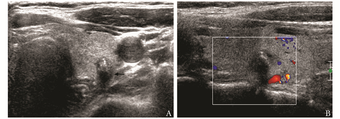
图 2 患者女,41岁,甲状腺超声示右叶中下部低回声0.7 cm×0.7 cm(箭头),形态不规则,边缘毛刺(A),彩色多普勒显像示血流信号局限性丰富杂乱(B),术后病理为甲状腺肿伴纤维化及钙化

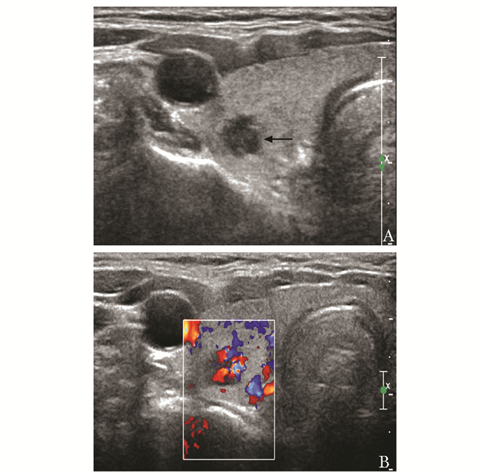
图 3 患者男,41岁,甲状腺超声示左叶中部背侧低回声0.7 cm×0.8 cm(箭头),边界欠清,纵横比大于1,内见微小钙化(A),彩色多普勒显像未见明显血流信号(B),术后病理为结节性甲状腺肿伴纤维化及钙化
表 1 33个被误诊的甲状腺良性病变病理结果及超声声像图特征
病理诊断 个数(%) 超声声像图特征(个) 极低回声 微小钙化 粗大钙化 边界不规则 纵横比≥1 血流异常 结节性甲状腺肿伴钙化/纤维化 12(36.36) 3 10 3 3 2 1 结节性甲状腺肿伴瘤样增生 7(21.21) 4 2 1 2 甲状腺滤泡性病变 6(18.18) 3 1 1 3 甲状腺炎 5(15.15) 1 2 2 甲状腺内纤维组织钙化 2(6.06) 1 1 1 陈旧性出血及纤维化 1(3.03) 1 1 总计 33(100) 3 20 5 9 6 6  下载: 导出CSV
-
[1] Cooper DS, Doherty GM, Haugen BR, et al. Revised American Thyroid Association Management Guidelines for patients with thyroid nodules and differentiated thyroid cancer[J].Thyroid, 2009, 19:1167-1264. doi: 10.1089/thy.2009.0110 [2] Cappelli C, Castellano M, Pirola I, et al.The predictive value of ultrasound findings in the management of thyroid nodules[J]. QJM, 2007, 100:29-35. http://www.wanfangdata.com.cn/details/detail.do?_type=perio&id=HighWire000001969009 [3] Salmaslioglu A, Erbil Y, Dural C, et al.Predictive value of sonographic features in preoperative evaluation of malignant thyroid nodules in a multinodular goiter[J].World J Surg, 2008, 32:1948-1954. doi: 10.1007/s00268-008-9600-2 [4] Horvath E, Majlis S, Rossi R, et al. An ultrasonogram reporting system for thyroid nodules stratifying cancer risk for clinical management[J]. J Clin Endocrinol Metab, 2009, 94:1748-1751. doi: 10.1210/jc.2008-1724 [5] 张益.高频彩超对甲状腺癌诊断价值的探讨和误诊分析[J].中国医师杂志, 2012, 14:402-403. http://www.wanfangdata.com.cn/details/detail.do?_type=perio&id=zgyszz201203044 [6] 刘术舟, 郭朱明.彩色多普勒超声在甲状腺癌中的诊断价值[J].中华普外科手术学杂志:电子版, 2013, 7:267-272. http://d.wanfangdata.com.cn/Periodical_zhptwksszz201304010.aspx [7] Cappelli C, Castellano M, Pirola I, et al.Thyroid nodule shape suggestes malignance[J].Eur J Endocrinol, 2006, 155:27-31. doi: 10.1530/eje.1.02177 [8] 张春梅, 吴长君, 张雪菊, 等.超声在甲状腺良恶性结节诊断中的应用[J].中国医学影像技术, 2007, 23:385-387. http://www.wanfangdata.com.cn/details/detail.do?_type=perio&id=zgyxyxjs200703019 [9] Das DK.Psammoma body:a product of dystrophic calcification or of a biologically active process that aims at limiting the growth and spread of tumor?[J].Diagn Cytopathol, 2009, 37:534-541. doi: 10.1002/dc.21081 [10] 刘丽, 徐辉雄, 吕明德, 等.甲状腺癌颈部淋巴结转移的超声特征[J].中华医学超声杂志:电子版, 2007, 4:156-158. http://d.wanfangdata.com.cn/Periodical/zhyxcszz200703011 [11] 洪玉蓉, 刘学明, 张秀芒, 等.超声检查甲状腺结节钙化类型与甲状腺肿癌的关系分析[J].中华超声影像学杂志, 2008, 17:977-980. http://www.cqvip.com/Main/Detail.aspx?id=28808925 [12] Kim SH, Kim BS, Jung SL, et al.Ultrasonographic findings of medullary thyroid carcinoma:a comparison with papillary thyroid carcinoma[J].Korean J Radiol, 2009, 10:101-105. doi: 10.3348/kjr.2009.10.2.101 [13] 尚卫国, 谭沛, 郭丹梅, 等.超声评估甲状腺结节纵横比≥ 1时对甲状腺微小癌的诊断价值[J].中国医师进修杂志, 2014, 37:59-61. http://www.wanfangdata.com.cn/details/detail.do?_type=perio&id=ysjxzz201427022 [14] 张华伟, 张志文, 梁波.二维及彩色多普勒超声对甲状腺乳头状癌的诊断价值[J].医学影像学杂志, 2012, 22:1661-1669. http://www.wanfangdata.com.cn/details/detail.do?_type=perio&id=yxyxxzz201210016 [15] 李泉水, 张家庭, 田平, 等.甲状腺癌的声像图特征研究[J].中国医学影像技术, 2006, 22:554-556. http://www.wanfangdata.com.cn/details/detail.do?_type=perio&id=zgyxyxjs200604023 [16] Salmaslioglu A, Erbil Y, Dural C, et al. Predicitive value of sonographic features in preoperative evaluation of malignant thyroid nodules in a multinodular goiter[J]. World J Surg, 2008, 32:1948-1954. doi: 10.1007/s00268-008-9600-2 [17] 刘术舟, 郭朱明.彩色多普勒超声在甲状腺癌中的诊断价值[J].中华普外科手术学杂志:电子版, 2013, 7:267-272. http://d.wanfangdata.com.cn/Periodical_zhptwksszz201304010.aspx [18] 吕珂, 姜玉新, 张缙熙, 等.甲状腺结节的超声诊断研究[J].中华超声影像学杂志, 2003, 12:285-288. http://www.wanfangdata.com.cn/details/detail.do?_type=perio&id=zhcsyx200305008 -

 点击查看大图
图(3) / 表(1)
计量
- 文章访问数: 201
- HTML全文浏览量: 70
- PDF下载量: 12
- 被引次数: 0


