 作者投稿
作者投稿 专家审稿
专家审稿 编辑办公
编辑办公 邮件订阅
邮件订阅 RSS
RSS

Comparison of Ultrasound and Magnetic Resonance Imaging in the Evaluation of Hand and Wrist Joint Involvement in Rheumatoid Arthritis
-
摘要:
目的 探讨类风湿关节炎腕手部关节病变的超声和磁共振病变分布特征并比较二者的诊断价值。 方法 对11例类风湿关节炎患者进行腕手部超声和磁共振检查, 计算两种影像方法对腕手部关节滑膜炎、骨质侵蚀和肌腱病变的检出率, 以磁共振为金标准评价超声对腕手部关节各种病变的诊断效力。 结果 共评价腕手部103个关节和112个肌腱区域。超声和磁共振对腕手部关节滑膜炎、骨质侵蚀、肌腱病变的检出率分别为59.2%、11.7%、18.8%和62.1%、14.6%、32.1%。腕关节三种病变检出率均高于掌指关节和指间关节。腕部伸肌腱病变检出率高于屈肌腱, 掌指关节屈肌腱病变检出率高于伸肌腱。与磁共振相比, 超声对腕手部关节滑膜炎、骨质侵蚀、肌腱病变的诊断敏感性为92.2%、73.3%、59.5%, 特异性为94.9%、97.7%、98.6%, 阳性预测值为96.7%、84.6%、96.2%, 阴性预测值为88.1%、95.6%、80.2%。 结论 类风湿关节炎腕部受累较掌指关节和近端指间关节常见, 腕部伸肌腱病变较屈肌腱病变常见, 掌指关节处屈肌腱病变较伸肌腱病变常见。以磁共振为对照, 超声检查对腕关节各种病变诊断准确率较高, 对掌指关节处伸肌腱病变诊断敏感性较低。 Abstract:Objective To compare the values of ultrasound and magnetic resonance imaging (MRI) in the diagnosis of hand and wrist joint involvement in rheumatoid arthritis(RA) patients and summarize the distribution pattern of these lesions. Methods A total of 11 RA cases were included. The detection rates of synovitis, bone erosion, and tenosynovitis of different joints were calculated based on ultrasound and MRI findings. The diagnostic efficiency of ultrasound was evaluated by using the MRI as golden standard. Results Totally 103 joints and 112 tendon areas were evaluated. The detection rates of synovitis, bone erosion, and tenosynovitis of wrist and hand joints were 59.2%, 11.7%, and 18.8% by ultrasound and 62.1%, 14.6%, and 32.1% by MRI. The detection rate of wrist joint was higher than metacarpophalangeal (MCP) or proximal interphalangeal(PIP) joints. The detection rate of extensor lesions was higher than that of flexor lesions at the wrist level; at the MCP level, however, the detection rate of flexor lesions was higher than that of extensor lesions. Compared with MRI, for the diagnosis of synovitis, bone erosion, and tenosynovitis, the ultrasound had a sensitivity of 92.2%, 73.3%, and 59.5%, a specificity of 94.9%, 97.7%, and 98.6%, a positive predictive value of 96.7%, 84.6%, and 96.2%, and a negative predictive value of 88.1%, 95.6%, and 80.2%. Conclusions Wrist joint involvement is more common than MCP and PIP joints involvement in RA patients. The tendon involvement is more common in extensor at the wrist level, while in flexor at the MCP level. Compared with MRI, the diagnostic efficiency of ultrasound is higher for wrist lesions than for hand joints lesions. However, ultrasound is less sensitive in the diagnosis of extensor lesions at the MCP level. -
Key words:
- ultrasound /
- magnetic resonance imaging /
- rheumatoid arthritis /
- joint involvement
-

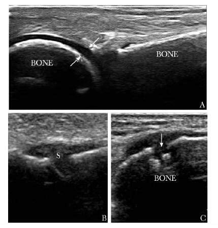
图 1 滑膜炎、骨质侵蚀的超声诊断标准
A.正常关节骨表面平滑连续,箭头所示为骨关节囊距离测量方法;B.滑膜增厚表现为骨关节囊距离增大,呈低回声;C.箭头所示为骨质侵蚀灶BONE:骨;S:滑膜炎
表 1 超声和磁共振对腕手部关节病变的检出率
病变 部位 关节数/例数 磁共振 超声 关节检出数/检出例数 百分比(%) 关节检出数/检出例数 百分比(%) 滑膜炎 W 22/11 17/11 77.3/100 15/10 68.2/90.9 MCP 45/9 23/6 51.1/66.7 24/6 53.3/66.7 PIP 36/9 24/7 66.7/77.8 22/7 61.1/77.8 合计 103/11 64/11 62.1/100 61/10 59.2/90.9 骨质侵蚀 W 22/11 12/8 54.5/72.7 10/7 45.5/63.6 MCP 45/9 2/2 4.4/22.2 1/1 2.2/11.1 PIP 36/9 1/1 2.8/11.1 1/1 2.8/11.1 合计 103/11 15/8 14.6/72.7 12/7 11.7/63.6 肌腱病变 W-F 11/11 5/5 45.5/45.5 4/4 36.4/36.4 W-E 11/11 8/8 72.7/72.7 7/7 63.6/63.6 MCP-F 45/9 18/6 40.0/66.7 9/4 20.0/44.4 MCP-E 45/9 7/4 15.6/44.4 1/1 2.2/11.1 合计 112/11 36/8 32.1/72.7 21/7 18.8/63.6 W:腕关节;MCP:掌指关节;PIP:近端指间关节;F:屈肌腱;E:伸肌腱  下载: 导出CSV
表 2 超声对腕手部关节滑膜炎、骨质侵蚀、肌腱病变的诊断效力(%)
病变 部位 敏感性 特异性 阳性预测值 阴性预测值 滑膜炎 W 82.4 80.0 93.3 57.1 MCP 100.0 95.5 95.8 100.0 PIP 91.7 100.0 100.0 85.7 合计 92.2 94.9 96.7 88.1 骨质侵蚀 W 75.0 12/8 90.0 75.0 MCP 50.0 97.7 50.0 97.7 PIP 100.0 100.0 100.0 100.0 合计 73.3 97.7 84.6 95.6 肌腱病变 W 84.6 100.0 100.0 81.8 MCP-F 50.0 100.0 100.0 75.0 MCP-E 14.3 100.0 100.0 86.4 合计 59.5 98.6 96.2 80.2 W、MCP、PIP、F、E:同表 1
下载: 导出CSV
-
[1] Schmidt WA, Schmidt H, Schicke B, et al. Standard reference values for musculoskeletal ultrasonography[J]. Ann Rheum Dis, 2004, 63:988-994. doi: 10.1136/ard.2003.015081 [2] Wakefield RJ, Balint PV, Szkudlarek M, et al. Musculoskeletal ultrasound including definitions for ultrasonographic pathology[J]. J Rheumatol, 2005, 32:24857. http://www.wanfangdata.com.cn/details/detail.do?_type=perio&id=f2029aec9f4b0081a3a8e220fa69d470 [3] 陈洋, 宗绍云, 李芹.超声在类风湿关节炎的应用及进展[J].医学影像学杂志, 2011, 21:767-770. http://www.wanfangdata.com.cn/details/detail.do?_type=perio&id=yxyxxzz201105045 [4] Tan YK, Østergaard M, Conaghan PG. Imaging tools in rheumatoid arthritis: ultrasound vsmagnetic resonance imaging[J]. Rheumatology, 2012, 51: vii36-vii42. [5] Mota LM, Laurindo IM, Neto LL, et al. Imaging diagnosis of early rheumatoid arthritis[J]. Rev Bras Reumatol, 2012, 52:757-766. http://europepmc.org/abstract/MED/23090375 [6] Rahmani M, Chegini H, Najafizadeh SR, et al. Detection of bone erosion in early rheumatoid arthritis:ultrasonography and conventional radiography versus non-contrast magnetic resonance imaging[J]. Clin Rheumatol, 2010, 29:88391. http://www.springerlink.com/content/b073135216h72781/ [7] Wakefield RJ, O'Connor PJ, Conaghan PG, et al. Finger tendon disease in untreated early rheumatoid arthritis:a comparison of ultrasound and magnetic resonance imaging[J]. Arthritis Rheum, 2007, 57:115864. [8] 王丽萍, 刘艳芳, 李应强, 等.类风湿性关节炎手部小关节病变的声像图表现[J].中华医学超声杂志:电子版, 2010, 7:450-455. http://www.wanfangdata.com.cn/details/detail.do?_type=perio&id=zhyxcszz201003015 [9] Ogishima H, Tsuboi H, Umeda N, et al. Analysis of subclinical synovitis detected by ultrasonographyand low-field magnetic resonance imaging in patients with rheumatoid arthritis[J]. Mod Rheumatol, 2014, 24:60-68. doi: 10.3109/14397595.2013.854050 [10] McQueen F, Beckley V, Crabbe J, et al. Magnetic resonance imaging evidence of tendinopathy in early rheumatoid arthritis predicts tendon rupture at six years[J]. Arthritis Rheum, 2005, 52:744-751. doi: 10.1002/art.20947 [11] Hoving JL, Buchbinder R, Hall S, et al. A comparison of magnetic resonance imaging, sonography, and radiography of the hand in patients with early rheumatoid arthritis[J]. J Rheumatol, 2004, 31:663-675. http://www.wanfangdata.com.cn/details/detail.do?_type=perio&id=976ad1c972e64c9a29de959f5a5338f7 -


 点击查看大图
图(2) / 表(2)
计量
- 文章访问数: 148
- HTML全文浏览量: 56
- PDF下载量: 13
- 被引次数: 0


