 作者投稿
作者投稿 专家审稿
专家审稿 编辑办公
编辑办公 邮件订阅
邮件订阅 RSS
RSS

Comparison of Prospective and Retrospective Electrocardiogram-triggered Coronary CT Angiography Using Dual-source CT
-
摘要:
目的 比较前瞻性心电触发序列扫描与回顾性心电触发螺旋扫描模式在双源CT冠状动脉成像中的图像质量及放射线剂量。 方法 将70例临床怀疑或已知冠心病的患者随机分为两组, 每组35例, 分别行前瞻性心电触发序列扫描和回顾性心电触发螺旋扫描冠状动脉CT成像, 对两种成像模式的图像质量及放射线剂量进行评价。 结果 两组患者的性别、年龄、体重指数匹配性良好, 差异无统计学意义(P > 0.05)。前瞻性心电触发序列扫描模式组和回顾性心电触发螺旋扫描模式组可评价的冠状动脉节段显示率分别为99.62%和99.62%, 两组间差异无统计学意义(χ2=0.000, P=1.000);两组图像质量评分分别为1.13±0.36和1.04±0.24, 差异有统计学意义(Z=-5.073, P=0.000);前瞻性心电触发序列扫描模式的放射线剂量为(3.47±1.00)mSv, 明显低于回顾性螺旋扫描模式的(14.28±1.81)mSv(P=0.032)。 结论 对于心律齐且心率≤ 70次/min的患者, 尽管前瞻性心电触发序列扫描的图像质量略差于回顾性螺旋扫描模式, 但两者可评价的冠状动脉节段显示率无明显差异, 而前者的有效放射剂量明显减少。 -
关键词:
- 冠状动脉成像 /
- X线计算机体层摄影术 /
- 影像质量 /
- 放射剂量
Abstract:Objective To compare the image quality and radiation exposure of prospective electrocardiogram (ECG) -triggered sequential and retrospective spiral acquisition coronary CT angiography by dual-source CT. Methods Seventy patients with suspected or known coronary artery disease were randomly divided into two groups (n=35). Group A underwent prospective ECG-triggered sequential scan and group B underwent retrospective ECG-triggered spiral scan. The image quality and radiation exposure of both modes were evaluated. Results There was no significant difference in gender, age, or body mass index between the two groups (P > 0.05). The rates of diagnostic coronary segments for group A and group B were 99.62% and 99.62%, respectively (χ2=0.000, P=1.000). The average image quality score was 1.13±0.36 in group A and 1.04±0.24 in group B, with significant difference between the two groups (Z=-5.073, P=0.000). The mean radiation dose of group A was significantly lower than that of group B[(3.47±1.00) mSv vs. (14.28±1.81) mSv, P=0.032]. Conclusion The prospective ECG-triggered sequential scan coronary CT angiography technique significantly reduces radiation dose without impairing the rates of diagnostic coronary segments when compared with the retrospective ECG-triggered spiral data acquisition in patients with a low and stable heart rate (≤ 70 bpm). -
Key words:
- coronary angiography /
- X-ray computed tomography /
- image quality /
- radiation exposure
-

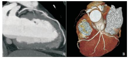
图 1 双源CT回顾性心电触发螺旋扫描冠状动脉成像
男,46岁,体重指数25. 7 kg /m2,心率53次/min,有效剂量14. 43 mSv,最大密度投影(A)及容积再现技术(B)显示前降支及其分支对角支,图像质量为1级,无搏动伪影

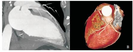
图 2 双源CT前瞻性心电触发序列扫描冠状动脉成像
男,57岁,体重指数23. 9 kg /m2,心率64次/min,有效剂量3. 53 mSv,最大密度投影(A)及容积再现技术(B)显示前降支及其分支对角支,可见阶梯状伪影,但血管连续,图像质量为2级
表 1 前瞻组与回顾组扫描参数的比较
组别 扫描方式 准直(mm) 采集层数(层) 机架旋转时间(s) 时间分辨率(ms) 管电压(kV) 管电流(mAs/转) 螺距 重建时相选择 前瞻组 序列扫描 32 × 0. 6 2 × 64 0. 33 83 120 371 无 70%时相 回顾组 螺旋扫描 32 × 0. 6 64 0. 33 83 120 400 0. 22 ~ 0. 33 35% ~ 75%时相  下载: 导出CSV
表 2 前瞻组与回顾组临床资料的比较(x ± s)
组别 性别(男/女) 年龄(岁) 体重指数(kg /m2) 扫描时心率(次/min) 扫描时间(s) 扫描时管电流(mAs/转) 前瞻组 19 /16 58. 7 ± 9. 1 24. 4 ± 2. 6 59. 9 ± 5. 8 0. 38 286. 06 ± 70. 69 回顾组 19 /16 59. 7 ± 11. 7 25. 6 ± 3. 1 61. 5 ± 4. 3 9. 98 ± 1. 12 400
下载: 导出CSV
表 3 前瞻组与回顾组放射剂量的比较(x ± s)
组别 CT容积剂量指数(mGy) 剂量长度乘积(mGy·cm) 有效放射剂量(mSv) 前瞻组 18. 45 ± 4. 55 247. 70 ± 71. 40 3. 47 ± 1. 00 回顾组 67. 78 ± 6. 64 1019. 77 ± 129. 27 14. 28 ± 1. 81 P值 0. 027 0. 032 0. 032
下载: 导出CSV
-
[1] Austen WG, Edwards JE, Frye RL, et al. A reporting system on patients evaluated for coronary artery disease:report of the Ad Hoc committee for grading of coronary artery disease, council on cardiovascular surgery[J]. Circulation, 1975, 51(4 Suppl):5-40. http://www.onacademic.com/detail/journal_1000040479488410_fa99.html [2] Stolzmann P, Leschka S, Scheffel H, et al. Dual-source CT in step-and-shoot mode:noninvasive coronary angiography with low radiation dose[J]. Radiology, 2008, 249:71-80. doi: 10.1148/radiol.2483072032 [3] Pflederer T, Jakstat J, Marwan M, et al. Radiation exposure and image quality in staged low dose protocols for coronary dual source CT angiography:a randomized comparison[J]. Eur Radiol, 2010, 20:1197-1206. doi: 10.1007/s00330-009-1645-6 [4] Hausleiter J, Meyer T, Hermann F, et al. Estimated radiation dose associated with cardiac CT angiography[J]. JAMA, 2009, 301:500-507. doi: 10.1001/jama.2009.54 [5] Pflederer T, Rudofsky L, Ropers D, et al. Image quality in a low radiation exposure protocol for retrospectively ECG-gated coronary CT angiography[J]. Am J Roentgenol, 2009, 192:1045-1050. doi: 10.2214/AJR.08.1025 [6] Scheffel H, Alkadhi H, Leschka S, et al. Low-dose CT coronary angiography in the step-and-shoot mode:diagnostic performance[J]. Heart, 2008, 94:1132-1137. doi: 10.1136/hrt.2008.149971 [7] Earls JP, Berman EL, Urban BA, et al. Prospectively gated transverse coronary CT angiography versus retrospectively gated helical technique:improved image quality and reduced radiation dose[J]. Radiology, 2008, 246:742-753. doi: 10.1148/radiol.2463070989 [8] Hirai N, Horiguchi J, Fujioka C, et al. Prospective versus retrospective ECG-gated 64-detector coronary CT angiography:assessment of image quality, stenosis, and radiation dose[J]. Radiology, 2008, 248:424-430. doi: 10.1148/radiol.2482071804 [9] Maruyama T, Takada M, Hasuike T, et al. Radiation dose reduction and coronary assessability of prospective electrocardiogram-gated computed tomography coronary angiography:comparison with retrospective electrocardiogramgated helical scan[J]. J Am Coll Cardiol, 2008, 52:1450-1455. doi: 10.1016/j.jacc.2008.07.048 [10] Efstathopoulos EP, Pantos I, Thalassinou S, et al. Patient radiation doses in cardiac computed tomography:comparison of published results with prospective and retrospective acquisition[J]. Radiat Prot Dosimetry, 2011, 1093:1-9. http://rpd.oxfordjournals.org/content/148/1/83 [11] Pontone G, Andreini D, Bartorelli AL, et al. Diagnostic accuracy of coronary computed tomography angiography:a comparison between prospective and retrospective electrocardiogram triggering[J]. J Am Coll Cardiol, 2009, 54:346-355. doi: 10.1016/j.jacc.2009.04.027 [12] 徐磊, 晏子旭, 张兆琪, 等.双源CT低剂量前瞻性心电触发序列扫描在冠状动脉血管成像的应用[J].中华放射学杂志, 2009, 43:700-703. http://www.wanfangdata.com.cn/details/detail.do?_type=perio&id=zhfsx200907008 [13] Shuman WP, Branch KR, May JM, et al. Prospective versus retrospective ECG gating for 64-detector CT of the coronary arteries:comparison of image quality and patient radiation dose[J]. Radiology, 2008, 248:431-437. doi: 10.1148/radiol.2482072192 -

 点击查看大图
图(2) / 表(3)
计量
- 文章访问数: 165
- HTML全文浏览量: 52
- PDF下载量: 8
- 被引次数: 0


