 作者投稿
作者投稿 专家审稿
专家审稿 编辑办公
编辑办公 邮件订阅
邮件订阅 RSS
RSS

-
摘要:
目的 探讨原发性肝血管肉瘤的病理诊断特点, 并复习相关文献。 方法 回顾性分析北京协和医院病理科2001年至2010年诊断的3例原发性肝血管肉瘤患者的病理及临床资料。 结果 3例患者的临床表现及影像所见无特异性, 镜下形态多样, 有血管瘤样、上皮样血管内皮细胞瘤样改变, 但均能找到较特异的血管肉瘤诊断特征。 结论 原发性肝血管肉瘤是一种罕见的恶性肿瘤, 形态复杂多变。病史、临床症状、影像检查、病理活检的综合运用, 对这一少见恶性肿瘤的诊断十分重要。 Abstract:Objective To explore the pathological manifestations of primary hepatic angiosarcoma and review the literature. Methods We retrospectively analyzed the pathological and clinical data of three cases of primary hepatic angiosarcoma, which were confirmed and treated in the Department of Pathology of Peking Union Medical College Hospital from 2001 to 2010. Results All these three cases showed no specific clinical manifestations and imaging results. Pathologically, there were varies of microscopic patterns such as hemangioma-like and epithelioid hemangioendothelioma-like apperieaces; however, specific pathological diagnostic features of angiosarcoma still existed in all three cases. Conclusions Primary hepatic angiosarcoma is a rare malignant tumor. The combined application of history-taking, symptom observation, imaging, and liver biopsy is important for the diagnosis of primary hepatic angiosarcoma. -
Key words:
- liver /
- angiosarcoma
-

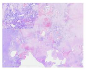
图 2 肿瘤区域弥漫出血充血背景下, 肝小叶结构消失, 散在灶性分布的增生小胆管间可见弥漫浸润性生长的新生毛细血管腔样结构及灶性密集排列的梭形细胞, 肿瘤周边浸润性生长; 高倍镜下可见小血管腔形成, 细胞异形性明显(HE, ×50, ×200)

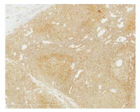
图 3 低倍镜下可见变性坏死及管腔形成, 呈海绵状血管瘤样改变, 部分区域细胞密集; 高倍镜下管腔内衬上皮及细胞密集区细胞异形性大, 可见奇异核(HE, ×10, ×200)
-
[1] Goodman ZD, Terraciano LM. Tumours and tumour-like lesions of the liver[M]//Mac Sween's Pathology of the Liver. 5th ed. Oxford: Churchill Livingstone, 2007: 761-814. [2] Ishak KG. Malignant mesenchymal tumours of the liver//Okuda K, Tabor E eds. Liver Cancer. Churchill Livingstone: New York, 1997. [3] Molina E, Hernandez A. Clinical manifestations primary hepatic angiosarcoma[J]. Dig Dis Sci, 2003, 48:677-682. doi: 10.1023/A:1022868221670 [4] Hamilton SR, Aaltonen LA. WHO pathology & genetics tumours of the digestive system[M]. 2000: 237-238. [5] Van Kampen RJ, Erdkamp FL, Peters FP. Thorium dioxiderelated hamangiosarcoma of the liver[J]. Neth J Med, 2007, 65:279-282. [6] Dogliotti E. Molecular mechanisms of carcinogenesis by vinyl chloride[J]. Ann 1st Super Sanita, 2006, 42:163-166. http://europepmc.org/abstract/MED/17033136 [7] Falk GW, Herbert J, Crowley S, et al. Epidemiology of hepatic angiosarcoma in the United States:1964-1974[J]. Environ Health Perspect, 1981, 41:107-113. doi: 10.1289/ehp.8141107 [8] Soini Y, Welsh JA, Ishak KG, et al. P53 mutation in primary hepatic angiosarcoma not associated with vinyl choride exposure[J]. Carcinogenesis, 1995, 16:2879-2881. doi: 10.1093/carcin/16.11.2879 [9] Przygodzki RM, Finkelstein SD, Keohavong P, et al. Sporadic and Thorotrast-induced angiosarcoma of the liver manifest frequent and multiple point mutations in K-ras-2[J]. Lab Invest, 1997, 76:153-159. http://www.wanfangdata.com.cn/details/detail.do?_type=perio&id=150a1ea41dc78f546b833f1d02584ca1 [10] Lincoln DT, Singal PK, Al-Banaw A. Growth hormone in vascular pathology:neovascularization and expression of receptors is associated with cellular proliferation[J]. Anticancer Res, 2007, 27:4201-4218. http://europepmc.org/abstract/MED/18225592 [11] Tate G, Suzuki T, Mitsuya T. Mutation of the PTEN gene in a human hepatic angiosarcoma[J]. Cancer Genet Cytogenet, 2007, 178:160-162. doi: 10.1016/j.cancergencyto.2007.07.017 [12] Koyama T, Fletcher JG, Johnson CD, et al. Primary hepatic angiosarcoma:findings at CT and MR Imaging[J]. Radiology, 2002, 222:667-673. doi: 10.1148/radiol.2223010877 [13] Weitz J, Klimstra DS, Cymes K, et al. Management of primary liver sarcoma[J]. Cancer, 2007, 109:1391-1396. doi: 10.1002/cncr.22530 [14] Lerut JP, Orlando G, Adam R, et al. The place of liver transplantation in the treatment of hepatic epitheloid hemangioendothelioma:report of the European liver transplant registry[J]. Ann Surg, 2007, 246:949-957. doi: 10.1097/SLA.0b013e31815c2a70 [15] Maluf D, Cotterell A, Clark B, et al. Hepatic angiosarcoma and liver transplantation:case report and literature review[J]. Transplant Proc, 2005, 37:2195-2199. doi: 10.1016/j.transproceed.2005.03.060 [16] Oe A, Habu D, Kawabe J, et al. A case of diffuse hepatic angiosarcoma diagnosed by FDG-PET[J]. Ann Nucl Med, 2005, 19:519-521. doi: 10.1007/BF02985582 [17] Shimada K, Nakamoto Y, Isoda H, et al. FDG PET for giant cavernous hemangioma:important clue to differentiate from a malignant vascular tumor in the liver[J]. Clin Nucl Med, 2010, 35:924-926. doi: 10.1097/RLU.0b013e3181f9de11 [18] Wang L, Lv K, Chang XY, et al. Contrast-enhanced ultrasound study of primary hepatic angiosarcoma: A pitfall of non-enhancement[J]. Eur J Radiol, 2011, Jul 6[Epub ahead of print]. PMID: 21737220. -

 下载:
下载:


 点击查看大图

图(5)
计量
- 文章访问数: 166
- HTML全文浏览量: 50
- PDF下载量: 1
- 被引次数: 0


