 作者投稿
作者投稿 专家审稿
专家审稿 编辑办公
编辑办公 邮件订阅
邮件订阅 RSS
RSS

Research Progress of Skin Tissue Engineering Scaffolds and Their Materials in Wound Repair
-
摘要: 近年来,皮肤组织工程技术为大面积创面修复提供了新的治疗思路。皮肤组织再生主要基于使用合适的组织支架,支架可细分为多孔、纤维、微球、水凝胶、脱细胞等几种类型,而支架材料又分为天然和合成两种,单一材料具有局限性,目前临床大多采用由两种及以上材料构成的复合材料。深入认识当前各种生物材料和支架的性能、优缺点,探索构建性能更佳的生物材料和支架,是未来皮肤组织工程研究的重要方向。本文对不同组织支架及支架材料在皮肤组织工程中的研究进展以及应用价值进行综述,以期为创面修复领域临床应用与研发提供参考。Abstract: In recent years, skin tissue engineering technology has provided new treatment ideas for large-area wound repair. Skin tissue regeneration is mainly based on the use of appropriate tissue scaffolds. There are several types of scaffolds, such as porous, fibrous, microsphere, hydrogel, and acellular scaffolds. Scaffold materials include natural materials and synthetic materials, but either type of single material has its own limitations. At present, most scaffolds are composed of two or more materials to form composites. Therefore an important direction of skin tissue engineering research in the future involves deeply understanding the properties, advantages and disadvantages of the various biomaterials and scaffolds currently used and exploring the construction of biomaterials and scaffolds with better performance. This article reviews the research progress and application value of different tissue scaffolds and scaffold materials in skin tissue engineering, in order to provide reference for clinical application and research and development in the field of wound repair.
-
Key words:
- scaffolds /
- biomaterials /
- synthetic /
- natural /
- polymers /
- skin /
- wound healing /
- tissue engineering
作者贡献:张思宇负责文献检索与论文撰写;马诗淇负责论文修订;王梦慈、李晓艺负责提取文献信息与整合;冯树梅提供写作思路、负责论文修订。利益冲突:所有作者均声明不存在利益冲突 -
表 2 合成生物材料及其优缺点
材料名称 优点 缺点 聚己内酯 具有较好的机械性能,降解缓慢,各种技术易于形成,水溶液中具备无收缩结构,成本低 疏水性,表面微观结构中无细胞识别位点[42] 聚乙烯吡咯烷酮 具有生物相容性[43] 具有超亲水性,伤口表面的水分可使其溶解[43] 聚乙二醇 植入后的免疫反应低,有助于在受伤后密封细胞膜,可限制细胞死亡[44] 细胞附着位点少 聚乳酸 可生物降解,具有生物相容性的聚合物,机械强度高,适合作为封装生物活性化合物的载体[45] 降解为酸性成分,导致局部酸度高,可能会破坏蛋白质,降解速度快 聚乳酸-羟基乙酸共聚物 无定形,可生物降解,具有与正常组织相似的特性 可能会经历塑性变形和失效  下载: 导出CSV
-
[1] Turner NJ, Badylak SF. The use of biologic scaffolds in the treatment of chronic nonhealing wounds[J]. Adv Wound Care, 2015, 4: 490-500. doi: 10.1089/wound.2014.0604 [2] Rezvani Ghomi E, Nourbakhsh N, Akbari Kenari M, et al. Collagen-based biomaterials for biomedical applications[J]. J Biomed Mater Res B Appl Biomater, 2021, 109: 1986-1999. doi: 10.1002/jbm.b.34881 [3] Troy E, Tilbury MA, Power AM, et al. Nature-based biomaterials and their application in biomedicine[J]. Polymers (Basel), 2021, 13: 3321. doi: 10.3390/polym13193321 [4] Park CH, Woo KM. Fibrin-based biomaterial applications in tissue engineering and regenerative medicine[J]. Adv Exp Med Biol, 2018, 1064: 253-261. http://www.ncbi.nlm.nih.gov/pubmed/30471038 [5] Wani SUD, Gautam SP, Qadrie ZL, et al. Silk fibroin as a natural polymeric based bio-material for tissue engineering and drug delivery systems-A review[J]. Int J Biol Macromol, 2020, 163: 2145-2161. doi: 10.1016/j.ijbiomac.2020.09.057 [6] Gholipourmalekabadi M, Sapru S, Samadikuchaksaraei A, et al. Silk fibroin for skin injury repair: Where do things stand?[J]. Adv Drug Deliv Rev, 2020, 153: 28-53. doi: 10.1016/j.addr.2019.09.003 [7] Zheng X, Chen Y, Dan N, et al. Highly stable collagen scaffolds crosslinked with an epoxidized natural polysac-charide for wound healing[J]. Int J Biol Macromol, 2021, 182: 1994-2002. doi: 10.1016/j.ijbiomac.2021.05.189 [8] Chandika P, Oh GW, Heo SY, et al. Electrospun porous bilayer nano-fibrous fish collagen/PCL bio-composite scaffolds with covalently cross-linked chitooligosaccharides for full-thickness wound-healing applications[J]. Mater Sci Eng C Mater Biol Appl, 2021, 121: 111871. doi: 10.1016/j.msec.2021.111871 [9] Tripathi S, Singh BN, Singh D, et al. Optimization and evaluation of ciprofloxacin-loaded collagen/chitosan scaffolds for skin tissue engineering[J]. 3 Biotech, 2021, 11: 160. doi: 10.1007/s13205-020-02567-w [10] Abolgheit S, Abdelkader S, Aboushelib M, et al. Bone marrow-derived mesenchymal stem cells and extracellular vesicles enriched collagen chitosan scaffold in skin wound healing (a rat model)[J]. J Biomater Appl, 2021, 36: 128-139. doi: 10.1177/0885328220963920 [11] Troy E, Tilbury M, Power A, et al. Nature-based biomaterials and their application in biomedicine[J]. Polymers (Basel), 2021, 13: 3321. doi: 10.3390/polym13193321 [12] Phull AR, Eo SH, Abbas Q, et al. Applications of Chondrocyte-Based Cartilage Engineering: An Overview[J]. Biomed Res Int, 2016, 2016: 1879837. http://www.onacademic.com/detail/journal_1000040476241210_6933.html [13] Su K, Wang C. Recent advances in the use of gelatin in biomedical research[J]. Biotechnol Lett, 2015, 37: 2139-2145. doi: 10.1007/s10529-015-1907-0 [14] Fang Q, Yao Z, Feng L, et al. Antibiotic-loaded chitosan-gelatin scaffolds for infected seawater immersion wound healing[J]. Int J Biol Macromol, 2020, 159: 1140-1155. doi: 10.1016/j.ijbiomac.2020.05.126 [15] Koyuncu A, Koç S, Akdere ÖE, et al. Investigation of the synergistic effect of platelet-rich plasma and polychromatic light on human dermal fibroblasts seeded chitosan/gelatin scaffolds for wound healing[J]. J Photochem Photobiol B, 2022, 232: 112476. doi: 10.1016/j.jphotobiol.2022.112476 [16] Al Kayal T, Losi P, Pierozzi S, et al. A new method for fibrin-based electrospun/sprayed scaffold fabrication[J]. Sci Rep, 2020, 10: 5111. doi: 10.1038/s41598-020-61933-z [17] Modery-Pawlowski CL, Tian LL, Pan V, et al. Approaches to synthetic platelet analogs[J]. Biomaterials, 2013, 34: 526-541. doi: 10.1016/j.biomaterials.2012.09.074 [18] Wong CC, Huang YM, Chen CH, et al. Cytokine and growth factor delivery from implanted platelet-rich fibrin enhances rabbit achilles tendon healing[J]. Int J Mol Sci, 2020, 21: 3221. doi: 10.3390/ijms21093221 [19] Suter N, Joshi A, Wunsch T, et al. Self-assembled fibrinogen nanofibers support fibroblast adhesion and prevent E. coli infiltration[J]. Mater Sci Eng C Mater Biol Appl, 2021, 126: 112156. doi: 10.1016/j.msec.2021.112156 [20] Sun W, Gregory DA, Tomeh MA, et al. Silk fibroin as a functional biomaterial for tissue engineering[J]. Int J Mol Sci, 2021, 22: 1499. doi: 10.3390/ijms22031499 [21] Fang G, Sapru S, Behera S, et al. Exploration of the tight structural-mechanical relationship in mulberry and non-mulberry silkworm silks[J]. J Mater Chem B, 2016, 4: 4337-4347. doi: 10.1039/C6TB01049K [22] Wu M, Huang S, Ye X, et al. Human epidermal growth factor-functionalized cocoon silk with improved cell proliferation activity for the fabrication of wound dressings[J]. J Biomater Appl, 2021, 36: 722-730. doi: 10.1177/0885328221997981 [23] Nguyen TP, Nguyen QV, Nguyen VH, et al. Silk fibroin-based biomaterials for biomedical applications: A review[J]. Polymers (Basel), 2019, 11: 1933. doi: 10.3390/polym11121933 [24] Cui B, Zhang C, Gan B, et al. Collagen-tussah silk fibroin hybrid scaffolds loaded with bone mesenchymal stem cells promote skin wound repair in rats[J]. Mater Sci Eng C Mater Biol Appl, 2020, 109: 110611. doi: 10.1016/j.msec.2019.110611 [25] Mogoᶊanu GD, Grumezescu AM. Natural and synthetic polymers for wounds and burns dressing[J]. Int J Pharm, 2014, 463: 127-136. doi: 10.1016/j.ijpharm.2013.12.015 [26] Yang MY, Liu BS, Huang HY, et al. Engineered pullulan-collagen-gold nano composite improves mesenchymal stem cells neural differentiation and inflammatory regulation[J]. Cells, 2021, 10: 3276. doi: 10.3390/cells10123276 [27] Dalgic AD, Koman E, Karatas A, et al. Natural origin bilayer pullulan-PHBV scaffold for wound healing applications[J]. Biomater Adv, 2022, 134: 112554. doi: 10.1016/j.msec.2021.112554 [28] Barbon S, Stocco E, Grandi F, et al. Biofabrication of a novel leukocyte-fibrin-platelet membrane as a cells and growth factors delivery platform for tissue engineering applications[J]. J Tissue Eng Regen Med, 2018, 12: 1891-1906. doi: 10.1002/term.2713 [29] Sharip NS, Ariffin H. Cellulose nanofibrils for biomaterial applications[J]. Mater Today Proc, 2019, 16: 1959-1968. doi: 10.1016/j.matpr.2019.06.074 [30] Hickey RJ, Pelling AE. Cellulose biomaterials for tissue engineering[J]. Front Bioeng Biotechnol, 2019, 7: 45. doi: 10.3389/fbioe.2019.00045 [31] Bar-Shai N, Sharabani-Yosef O, Zollmann M, et al. Seaweed cellulose scaffolds derived from green macroalgae for tissue engineering[J]. Sci Rep, 2021, 11: 11843. doi: 10.1038/s41598-021-90903-2 [32] Subhedar A, Bhadauria S, Ahankari S, et al. Nanocellulose in biomedical and biosensing applications: A review[J]. Int J Biol Macromol, 2021, 166: 587-600. doi: 10.1016/j.ijbiomac.2020.10.217 [33] Fu L, Zhou P, Zhang S, et al. Evaluation of bacterial nanocellulose-based uniform wound dressing for large area skin transplantation[J]. Mater Sci Eng C Mater Biol Appl, 2013, 33: 2995-3000. doi: 10.1016/j.msec.2013.03.026 [34] Yang G, Xie J, Hong F, et al. Antimicrobial activity of silver nanoparticle impregnated bacterial cellulose membr-ane: Effect of fermentation carbon sources of bacterial cellulose[J]. Carbohydr Polym, 2012, 87: 839-845. doi: 10.1016/j.carbpol.2011.08.079 [35] Younes I, Rinaudo M. Chitin and chitosan preparation from marine sources. Structure, properties and applications[J]. Mar Drugs, 2015, 13: 1133-1174. doi: 10.3390/md13031133 [36] Rezaei FS, Sharifianjazi F, Esmaeilkhanian A, et al. Chitosan films and scaffolds for regenerative medicine applications: A review[J]. Carbohydr Polym, 2021, 273: 118631. doi: 10.1016/j.carbpol.2021.118631 [37] Zhao H, Liao J, Wu F, et al. Mechanical strength improvement of chitosan/hydroxyapatite scaffolds by coating and cross-linking[J]. J Mech Behav Biomed Mater, 2021, 114: 104169. doi: 10.1016/j.jmbbm.2020.104169 [38] Hu Z, Zhang D, Lu S, et al. Chitosan-based composite materials for prospective hemostatic applications[J]. Mar Drugs, 2018, 16: 273. doi: 10.3390/md16080273 [39] Jiang Z, Zhang K, Du L, et al. Construction of chitosan scaffolds with controllable microchannel for tissue engineer-ing and regenerative medicine[J]. Mater Sci Eng C Mater Biol Appl, 2021, 126: 112178. doi: 10.1016/j.msec.2021.112178 [40] Salesa B, Llorens-Gámez M, Serrano-Aroca Á. Study of 1D and 2D carbon nanomaterial in alginate films[J]. Nanomaterials (Basel), 2020, 10: 206. doi: 10.3390/nano10020206 [41] Piras CC, Smith DK. Multicomponent polysaccharide alginate-based bioinks[J]. J Mater Chem B, 2020, 8: 8171-8188. doi: 10.1039/D0TB01005G [42] Ghosal K, Manakhov A, Zajíčková L, et al. Structural and surface compatibility study of modified electrospun poly(ε-caprolactone) (PCL) composites for skin tissue engineering[J]. AAPS Pharm Sci Tech, 2017, 18: 72-81. doi: 10.1208/s12249-016-0500-8 [43] Hajikhani M, Emam-Djomeh Z, Askari G. Fabrication and characterization of mucoadhesive bioplastic patch via coaxial polylactic acid (PLA) based electrospun nanofibers with antimicrobial and wound healing application[J]. Int J Biol Macromol, 2021, 172: 143-153. doi: 10.1016/j.ijbiomac.2021.01.051 [44] Cheng CC, Yang XJ, Fan WL, et al. Cytosine-Functiona-lized Supramolecular Polymer-Mediated Cellular Behavior and Wound Healing[J]. Biomacromolecules, 2020, 21: 3857-3866. doi: 10.1021/acs.biomac.0c00938 [45] Farah S, Anderson DG, Langer R. Physical and mechanical properties of PLA, and their functions in widespread applications — A comprehensive review[J]. Adv Drug Deliv Rev, 2016, 107: 367-392. doi: 10.1016/j.addr.2016.06.012 [46] Sadeghi-Avalshahr AR, Nokhasteh S, Molavi A, et al. Tailored PCL scaffolds as skin substitutes using sacrificial PVP fibers and collagen/chitosan blends[J]. Int J Mol Sci, 2020, 21: 2311. doi: 10.3390/ijms21072311 [47] Liu C, Wang Z, Wei X, et al. 3D printed hydrogel/PCL core/shell fiber scaffolds with NIR-triggered drug release for cancer therapy and wound healing[J]. Acta Biomater, 2021, 131: 314-325. doi: 10.1016/j.actbio.2021.07.011 [48] Lu H, Oh HH, Kawazoe N, et al. PLLA-collagen and PLLA-gelatin hybrid scaffolds with funnel-like porous structure for skin tissue engineering[J]. Sci Technol Adv Mater, 2012, 13: 064210. doi: 10.1088/1468-6996/13/6/064210 [49] Panayi AC, Haug V, Liu Q, et al. Novel application of autologous micrografts in a collagen-glycosaminoglycan scaffold for diabetic wound healing[J]. Biomed Mater, 2021, 16: 035032. doi: 10.1088/1748-605X/abc3dc [50] Vasita R, Katti DS. Nanofibers and their applications in tissue engineering[J]. Int J Nanomed, 2006, 1: 15-30. doi: 10.2147/nano.2006.1.1.15 [51] Zahedi E, Esmaeili A, Eslahi N, et al. Fabrication and Characterization of Core-Shell Electrospun Fibrous Mats Containing Medicinal Herbs for Wound Healing and Skin Tissue Engineering[J]. Mar Drugs, 2019, 17: 27. doi: 10.3390/md17010027 [52] Elliott WH, Bonani W, Maniglio D, et al. Silk hydrogels of tunable structure and viscoelastic properties using different chronological orders of genipin and physical cross-linking[J]. ACS Appl Mater Interfaces, 2015, 7: 12099-12108. doi: 10.1021/acsami.5b02308 [53] Shafei S, Khanmohammadi M, Heidari R, et al. Exosome loaded alginate hydrogel promotes tissue regeneration in full-thickness skin wounds: An in vivo study[J]. J Biomed Mater Res A, 2020, 108: 545-556. doi: 10.1002/jbm.a.36835 [54] Muñoz-González PU, Lona-Ramos MC, Gutiérrez-Verdín LD, et al. Gel dressing based on type Ⅰ collagen modified with oligourethane and silica for skin wound healing[J]. Biomed Mater, 2022, 17: 045005. doi: 10.1088/1748-605X/ac6b70 [55] Lee S, Choi E, Cha MJ, et al. Cell adhesion and long-term survival of transplanted mesenchymal stem cells: a prerequisite for cell therapy[J]. Oxid Med Cell Longev, 2015, 2015: 632902. doi: 10.1155/2015/632902 [56] Zhang Z, Li Z, Li Y, et al. Sodium alginate/collagen hydrogel loaded with human umbilical cord mesenchymal stem cells promotes wound healing and skin remodeling[J]. Cell Tissue Res, 2021, 383: 809-821. doi: 10.1007/s00441-020-03321-7 [57] Solarte David VA, Güiza-Argüello VR, Arango-Rodríguez ML, et al. Decellularized Tissues for Wound Healing: Towards Closing the Gap Between Scaffold Design and Effective Extracellular Matrix Remodeling[J]. Front Bioeng Biotechnol, 2022, 10: 821852. doi: 10.3389/fbioe.2022.821852 [58] Ahmed Omar N, Amédée J, Letourneur D, et al. Recent advances of pullulan and/or dextran-based materials for bone tissue engineering strategies in preclinical studies: A systematic review[J]. Front Bioeng Biotechnol, 2022, 10: 889481. doi: 10.3389/fbioe.2022.889481 [59] Nyame TT, Chiang HA, Leavitt T, et al. Tissue-engineered skin substitutes[J]. Plast Reconstr Surg, 2015, 136: 1379-1388. doi: 10.1097/PRS.0000000000001748 [60] Wang F, Wang M, She Z, et al. Collagen/chitosan based two-compartment and bi-functional dermal scaffolds for skin regeneration[J]. Mater Sci Eng C Mater Biol Appl, 2015, 52: 155-162. doi: 10.1016/j.msec.2015.03.013 [61] Lastra ML, Gómez Ribelles JL, Cortizo AM. Design and characterization of microspheres for a 3D mesenchymal stem cell culture[J]. Colloids Surf B Biointerfaces, 2020, 196: 111322. doi: 10.1016/j.colsurfb.2020.111322 [62] Huang S, Lu G, Wu Y, et al. Mesenchymal stem cells delivered in a microsphere-based engineered skin contribute to cutaneous wound healing and sweat gland repair[J]. J Dermatol Sci, 2012, 66: 29-36. doi: 10.1016/j.jdermsci.2012.02.002 -

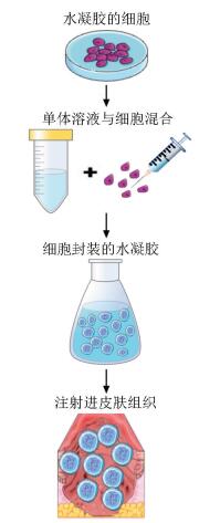
 点击查看大图
图(1) / 表(2)
计量
- 文章访问数: 551
- HTML全文浏览量: 84
- PDF下载量: 75
- 被引次数: 0


