 作者投稿
作者投稿 专家审稿
专家审稿 编辑办公
编辑办公 邮件订阅
邮件订阅 RSS
RSS

Novel Real-time MRI Navigation Technology in the Treatment of Venous Malformation: A Case Report
-
摘要: 静脉畸形是一种常见的先天性血管发育异常, 血管内硬化治疗是其治疗的一线方案。对病灶进行准确穿刺是血管内硬化治疗的关键, 如穿刺有偏差, 很可能造成局部溃疡、坏死等并发症。本例静脉畸形患者在诊疗中应用了新研发的实时MRI导航技术, 顺利实现了对常规手段难以到达的深部病变进行硬化剂注射治疗的目标。本文通过对该病例的诊疗经过进行回顾, 以期为静脉畸形或其他软组织疾病的诊断与治疗提供借鉴。Abstract: Venous malformation is a common congenital anomaly of vascular development, and endovascular sclerotherapy is currently the first-line treatment for it. Accurate puncture of the lesion is the key to endovascular treatment. Any deviation is likely to result in local ulcers, necrosis and other complications. We described one case, in which a newly developed real-time MRI navigation technology was introduced and sclerotherapy was successfully performed for deep lesions that were difficult to reach by conventional methods. This article reviewed the treatment process of this case, in order to provide guidance and help for venous malformations and other soft tissue diseases in the future.作者贡献:周经纬、张紫旻负责收集资料,撰写论文;顾豪协助疏理撰稿思路;陈辉、胡丽负责提供技术支持,审阅论文;刘泓源、徐梓安负责收集、核对资料;杨希、林晓曦负责指导论文修订。利益冲突:所有作者均声明不存在利益冲突
-
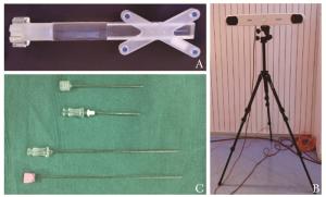
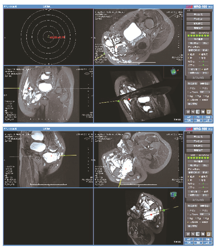
图 2 实时MRI导航系统主要定位设备及穿刺针
A.可拆卸式床旁定位架,可根据治疗部位安装在检查床的不同位置; B.红外摄像仪可捕捉追踪标记反射的光线以推算器械、检查床和MRI设备的相对位置; C.18G/5 cm和18G/10 cm两种型号的磁兼容钛合金穿刺针
-
[1] Behravesh S, Yakes W, Gupta N, et al. Venous malformations: clinical diagnosis and treatment[J]. Cardiovasc Diagn Ther, 2016, 6: 557-569. doi: 10.21037/cdt.2016.11.10 [2] Richter GT, Friedman AB. Hemangiomas and vascular malformations: current theory and management[J]. Int J Pediatr, 2012, 2012: 645678. [3] Cooke-Barber J, Kreimer S, Patel M, et al. Venous malformations[J]. Semin Pediatr Surg, 2020, 29: 150976. doi: 10.1016/j.sempedsurg.2020.150976 [4] Carqueja IM, Sousa J, Mansilha A. Vascular malformations: classification, diagnosis and treatment[J]. Int Angiol, 2018, 37: 127-142. [5] Hage AN, Chick JFB, Srinivasa RN, et al. Treatment of Venous Malformations: The Data, Where We Are, and How It Is Done[J]. Tech Vasc Interv Radiol, 2018, 21: 45-54. doi: 10.1053/j.tvir.2018.03.001 [6] Mulligan PR, Prajapati HJS, Martin LG, et al. Vascular anomalies: classification, imaging characteristics and implications for interventional radiology treatment approaches[J]. Br J Radiol, 2014, 87: 20130392. doi: 10.1259/bjr.20130392 [7] Song D, Guo L, Sheng H, et al. DSA-guided percutaneous sclerotherapy for children with oropharyngeal low-flow venous malformation[J]. Exp Ther Med, 2020, 19: 3405-3410. [8] Song D, Wu C, Guo L, et al. Efficacy and safety of DSA-guided percutaneous sclerotherapy for venous malformations of penile region in children[J]. J Pediatr Surg, 2021, 56: 601-604. doi: 10.1016/j.jpedsurg.2020.07.020 [9] Vollherbst DF, Gebhart P, Kargus S, et al. Image-guided percutaneous sclerotherapy of venous malformations of the head and neck: Clinical and MR-based volumetric mid-term outcome[J]. PLoS One, 2020, 15: e0241347. doi: 10.1371/journal.pone.0241347 [10] Kumar S, Bhavana K, Kumar S, et al. Ultrasound-guided polidocanol foam sclerotherapy for treating venous malformations[J]. J Clin Ultrasound, 2018, 46: 23-31. doi: 10.1002/jcu.22546 [11] Goldman LH, Perronne L, Alaia EF, et al. Does Magnetic Resonance Imaging After Diagnostic Ultrasound for Soft Tissue Masses Change Clinical Management[J]. J Ultrasound Med, 2021, 40: 1515-1522. doi: 10.1002/jum.15529 [12] Clemens RK, Baumann F, Husmann M, et al. Percutaneous sclerotherapy for spongiform venous malformations-analysis of patient-evaluated outcome and satisfaction[J]. Vasa, 2017, 46: 477-483. doi: 10.1024/0301-1526/a000650 [13] 中华医学会整形外科分会血管瘤和脉管畸形学组. 血管瘤和脉管畸形的诊断及治疗指南(2019版)[J]. 组织工程与重建外科杂志, 2019, 15: 277-317. doi: 10.3969/j.issn.1673-0364.2019.05.001 [14] Navarro OM, Laffan EE, Ngan BY. Pediatric Soft-Tissue Tumors and Pseudo-tumors: MR Imaging Features with Pathologic Correlation[J]. Radio Graphics, 2009, 29: 887-906. [15] Navarro OM. Magnetic resonance imaging of pediatric soft-tissue vascular anomalies[J]. Pediatr Radiol, 2016, 46: 891-901. doi: 10.1007/s00247-016-3567-1 [16] Fayad L, Hazirolan T, Bluemke D, et al. Vascular malformations in the extremities: emphasis on MR imaging features that guide treatment options[J]. Skeletal Radiol, 2006, 35: 127-137. doi: 10.1007/s00256-005-0057-1 [17] Andreisek G, Nanz D, Weishaupt D, et al. MR imaging-guided percutaneous sclerotherapy of peripheral venous malformations with a clinical 1.5-T unit: a pilot study[J]. J Vasc Interv Radiol, 2009, 20: 879-887. doi: 10.1016/j.jvir.2009.03.034 [18] Partovi S, Lu Z, Vidal L, et al. Real-time MRI-guided percutaneous sclerotherapy treatment of venous low-flow malformations in the head and neck[J]. Phlebology, 2018, 33: 344-352. doi: 10.1177/0268355517710110 [19] Partovi S, Vidal L, Lu Z, Nakamoto DA, et al. Real-time MRI-guided percutaneous sclerotherapy of low-flow head and neck lymphatic malformations in the pediatric population-a stepwise approach[J]. Pediatr Radiol, 2017, 47: 755-760. doi: 10.1007/s00247-017-3789-x [20] Rothgang E, Gilson WD, Wacker F, et al. Rapid freehand MR-guided percutaneous needle interventions: an image-based approach to improve workflow and feasibility[J]. J Magn Reson Imaging, 2013, 37: 1202-1212. doi: 10.1002/jmri.23894 [21] O'Mara DM, DiCamillo PA, Gilson WD, et al. MR-guided percutaneous sclerotherapy of low-flow vascular malformations: Clinical experience using a 1.5 tesla MR system[J]. J Magn Reson Imaging, 2017, 45: 1154-1162. doi: 10.1002/jmri.25502 [22] O'Mara DM, Berges AJ, Fritz J, et al. MRI-guided percutaneous sclerotherapy of venous malformations: initial clinical experience using a 3T MRI system[J]. Clin Imaging, 2020, 65: 8-14. doi: 10.1016/j.clinimag.2020.04.012 -

 下载:
下载:


 点击查看大图
图(5)
计量
- 文章访问数: 455
- HTML全文浏览量: 70
- PDF下载量: 32
- 被引次数: 0


